Image Library

Searchable collection of capillaroscopic images organized by pattern, diagnosis, and severity.

Capillaroscopy Image Atlas

Image Library

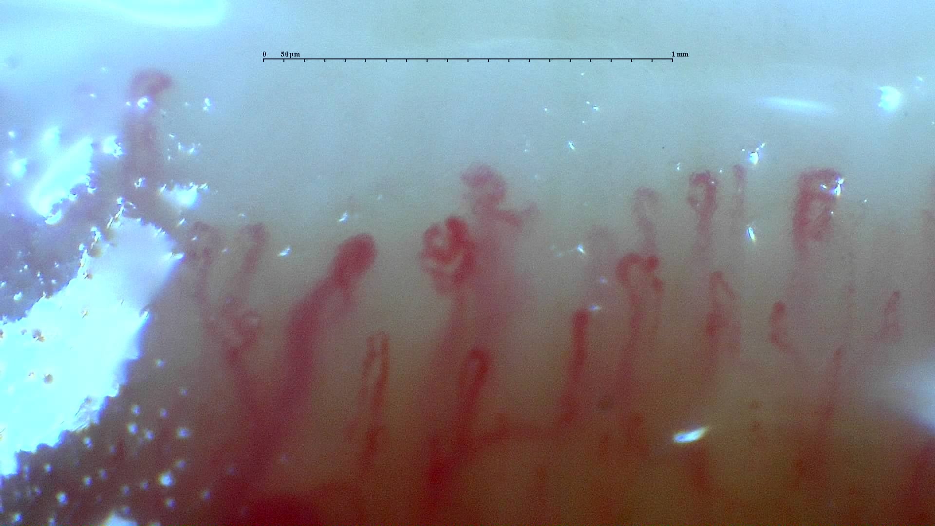
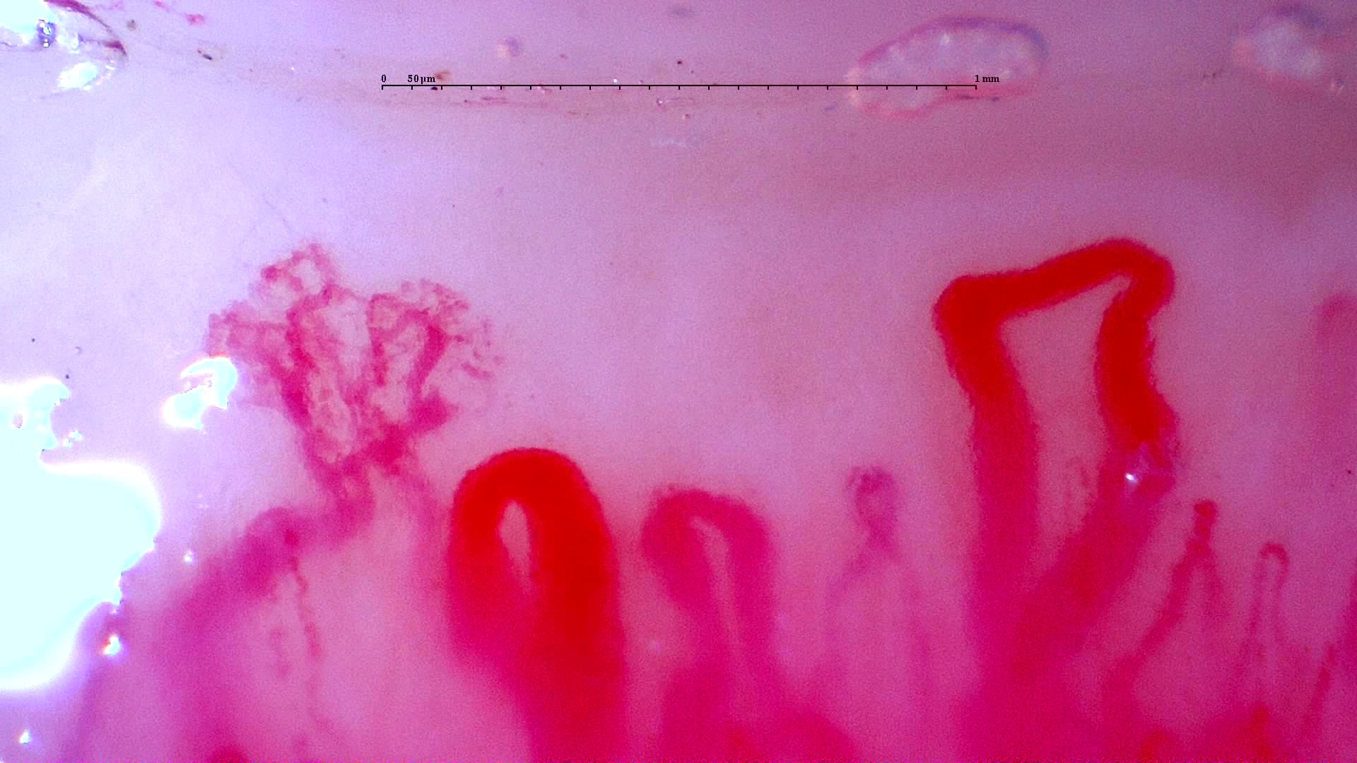
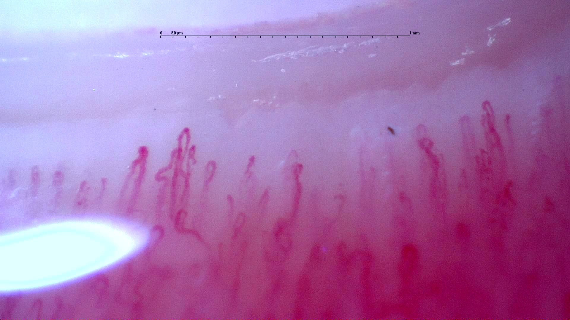
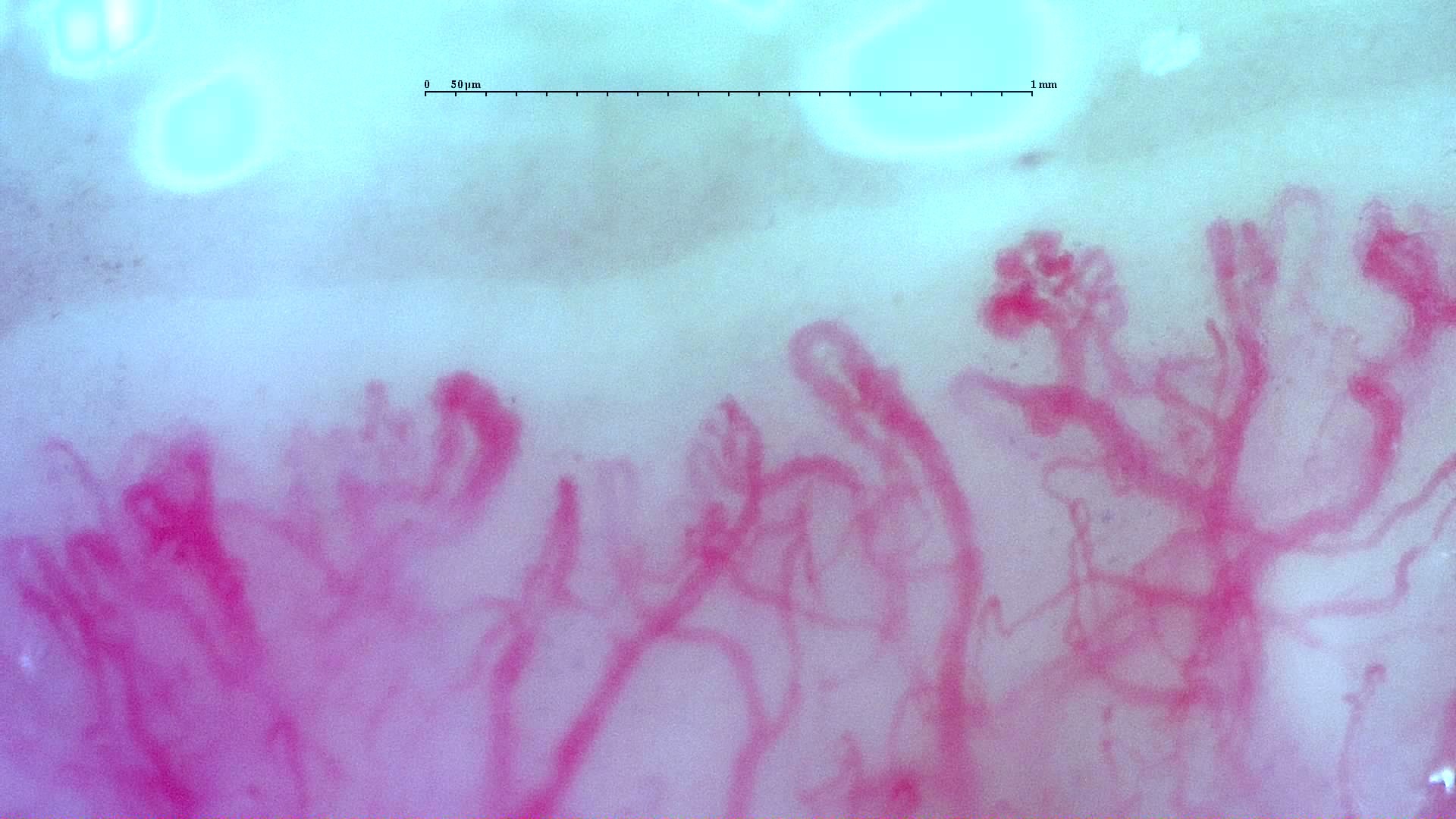
This image library is designed as a structured atlas of key capillaroscopic hallmarks, patterns, and pathology-associated findings to support fast visual learning and case comparison.

Total images: 43Pathologies: 21Patterns: 29
Filters

Click tags to filter images. Click again to remove a tag.

Pathology

Severity

Pattern

Hallmarks

Showing 43 of 43 images

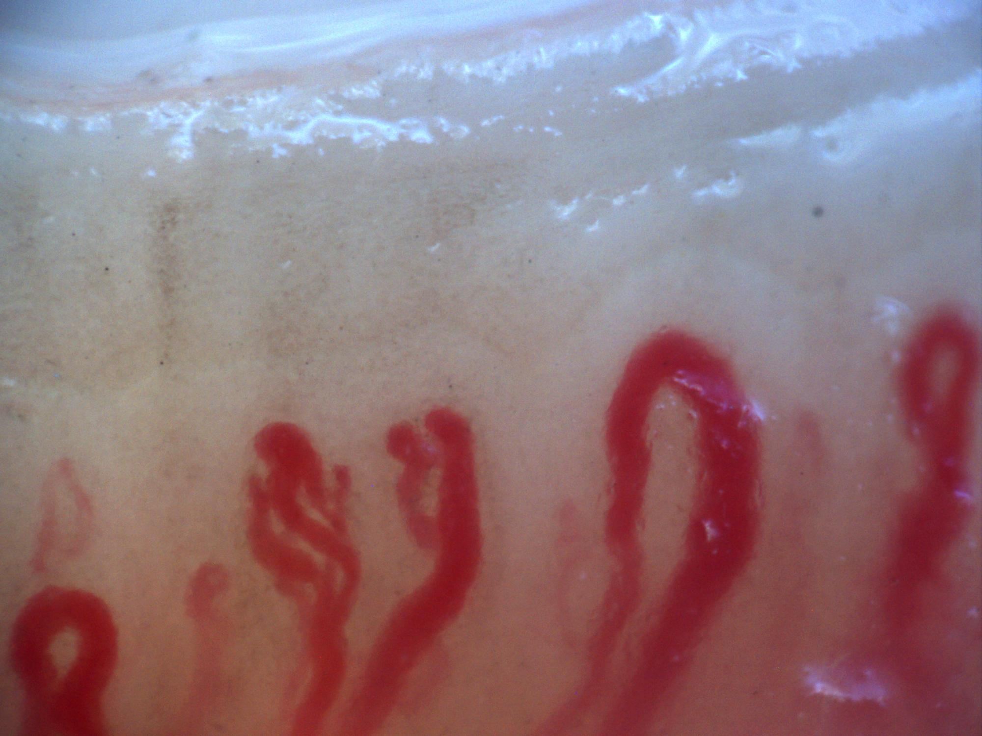
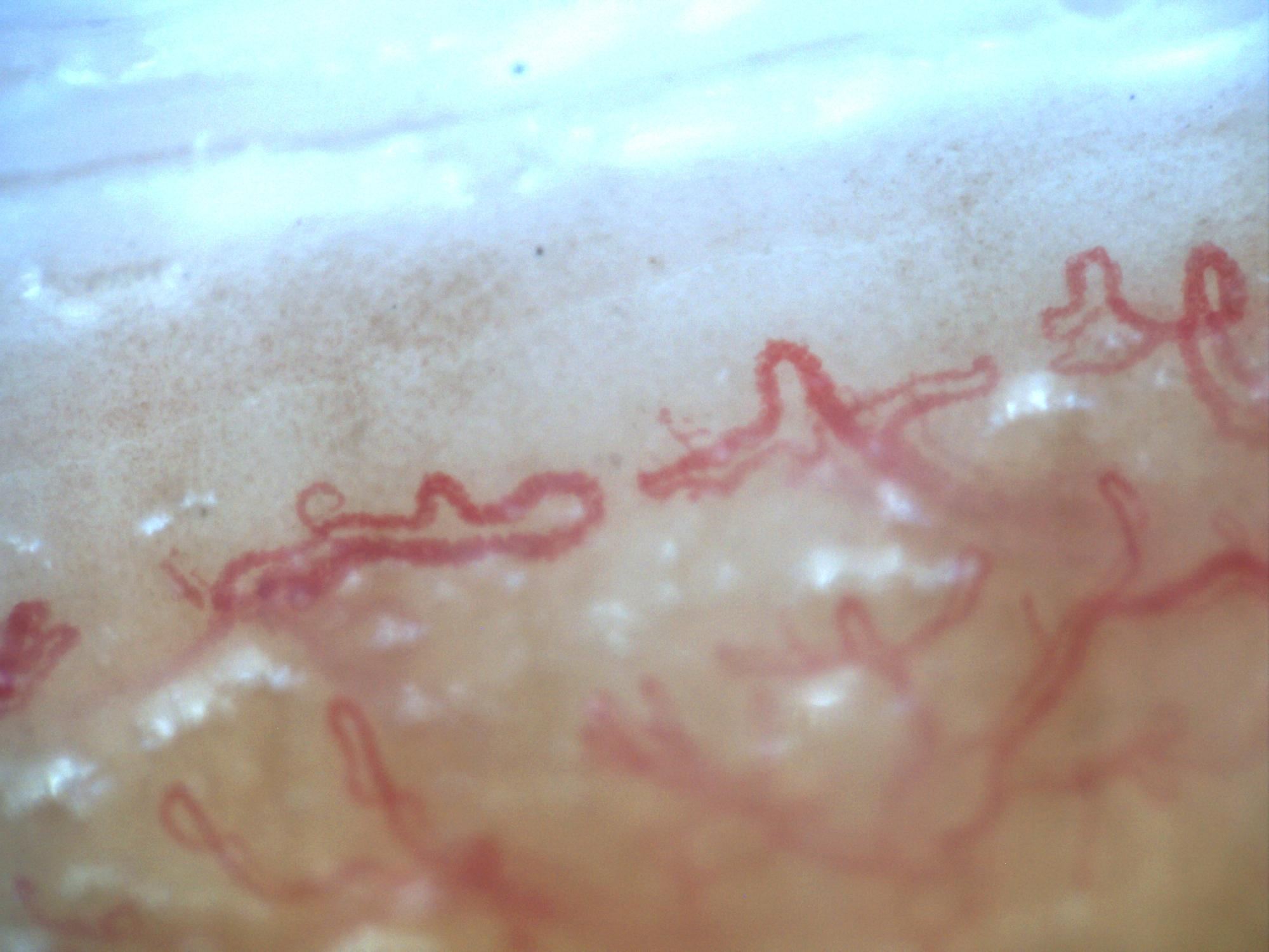
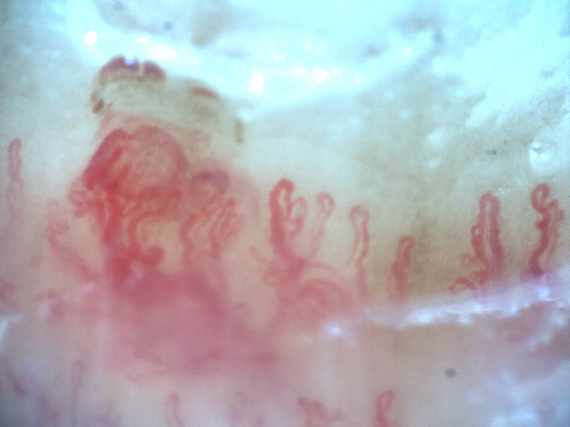
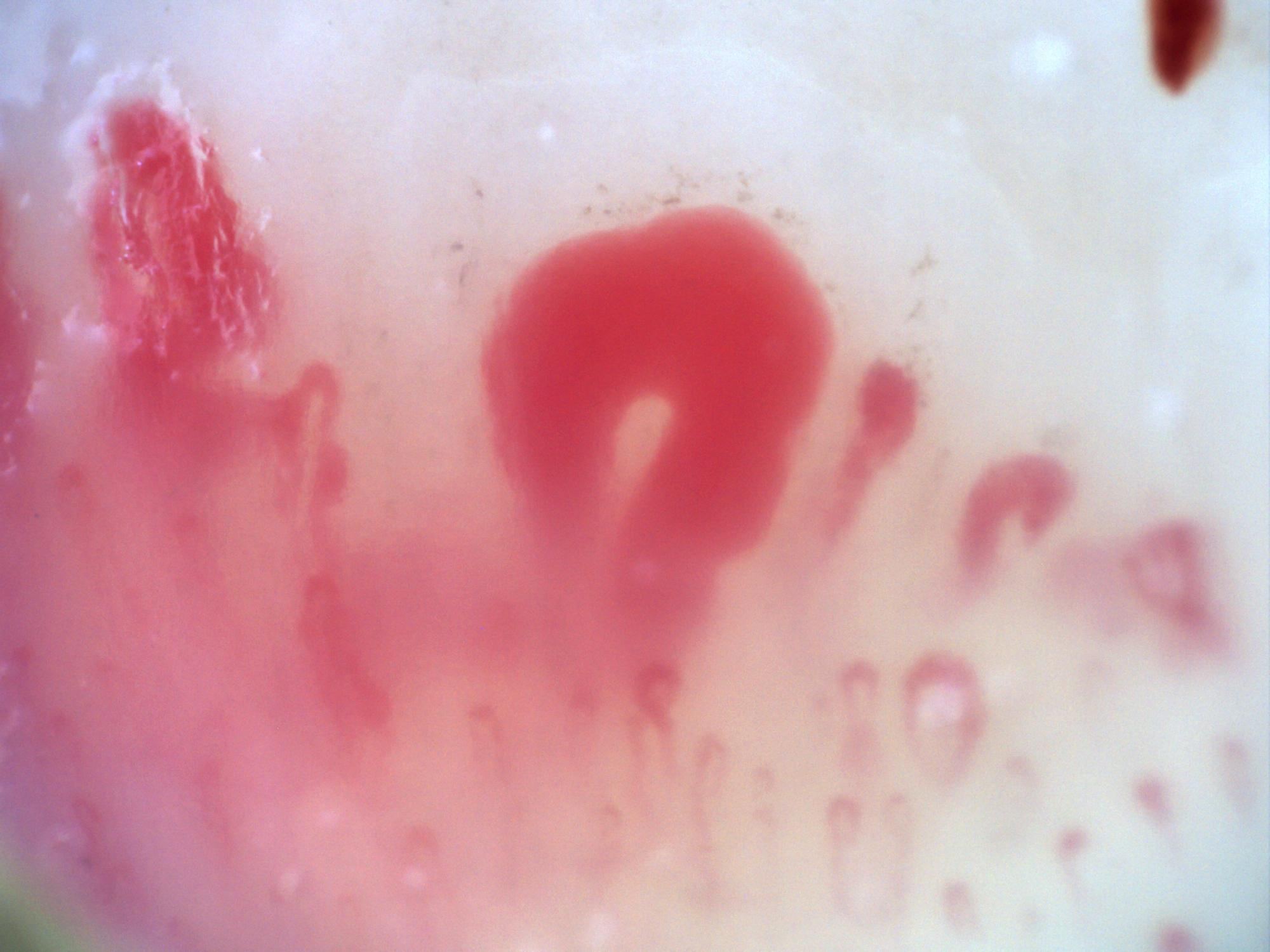
200x nailfold capillaroscopy field with several megacapillaries in an active scleroderma pattern
View details

Active scleroderma pattern with megacapillaries (field 1)

Author: Miguel Antonio Mesa Navas

Systemic sclerosisActive scleroderma patternModerate to Severe
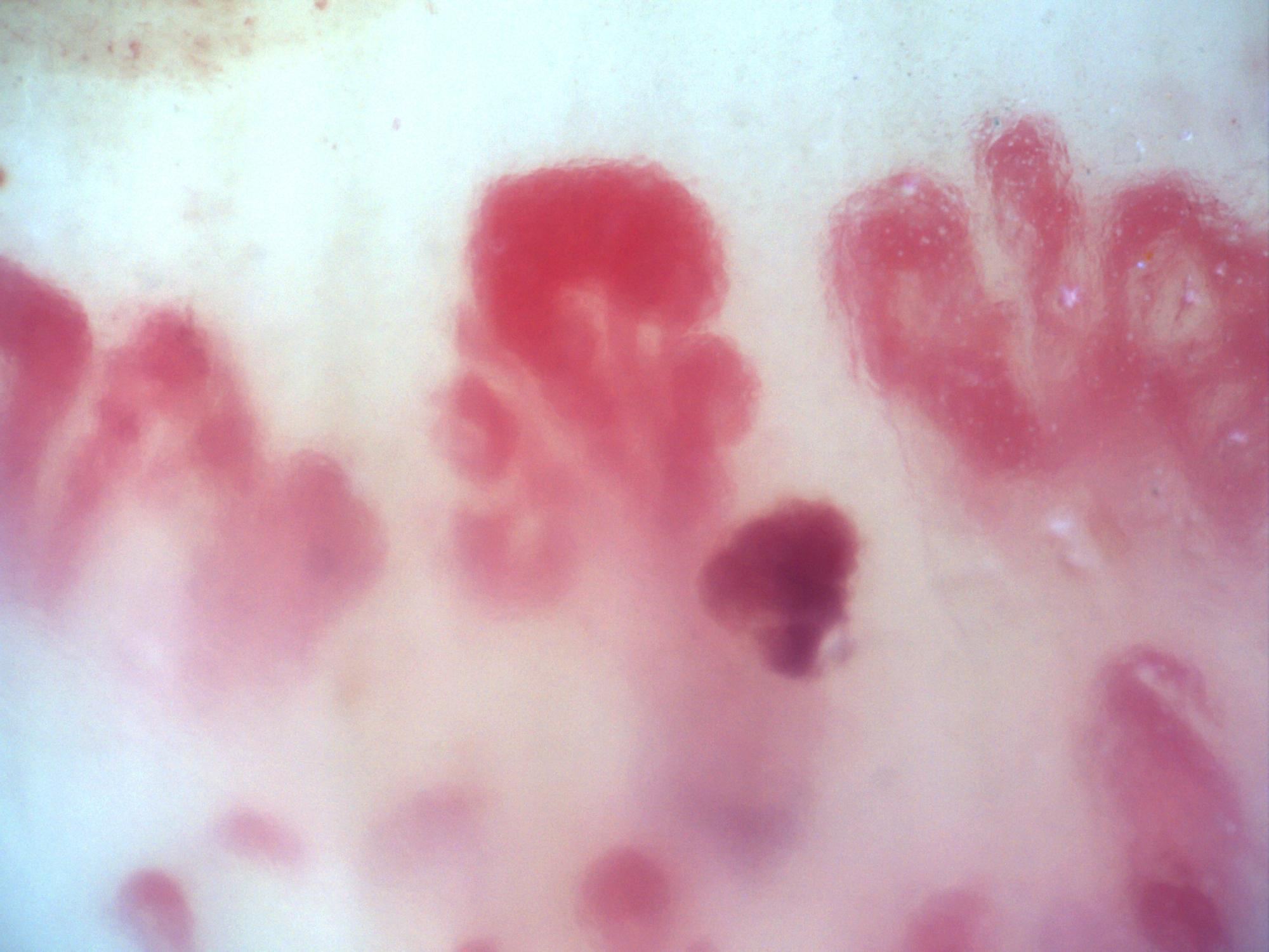
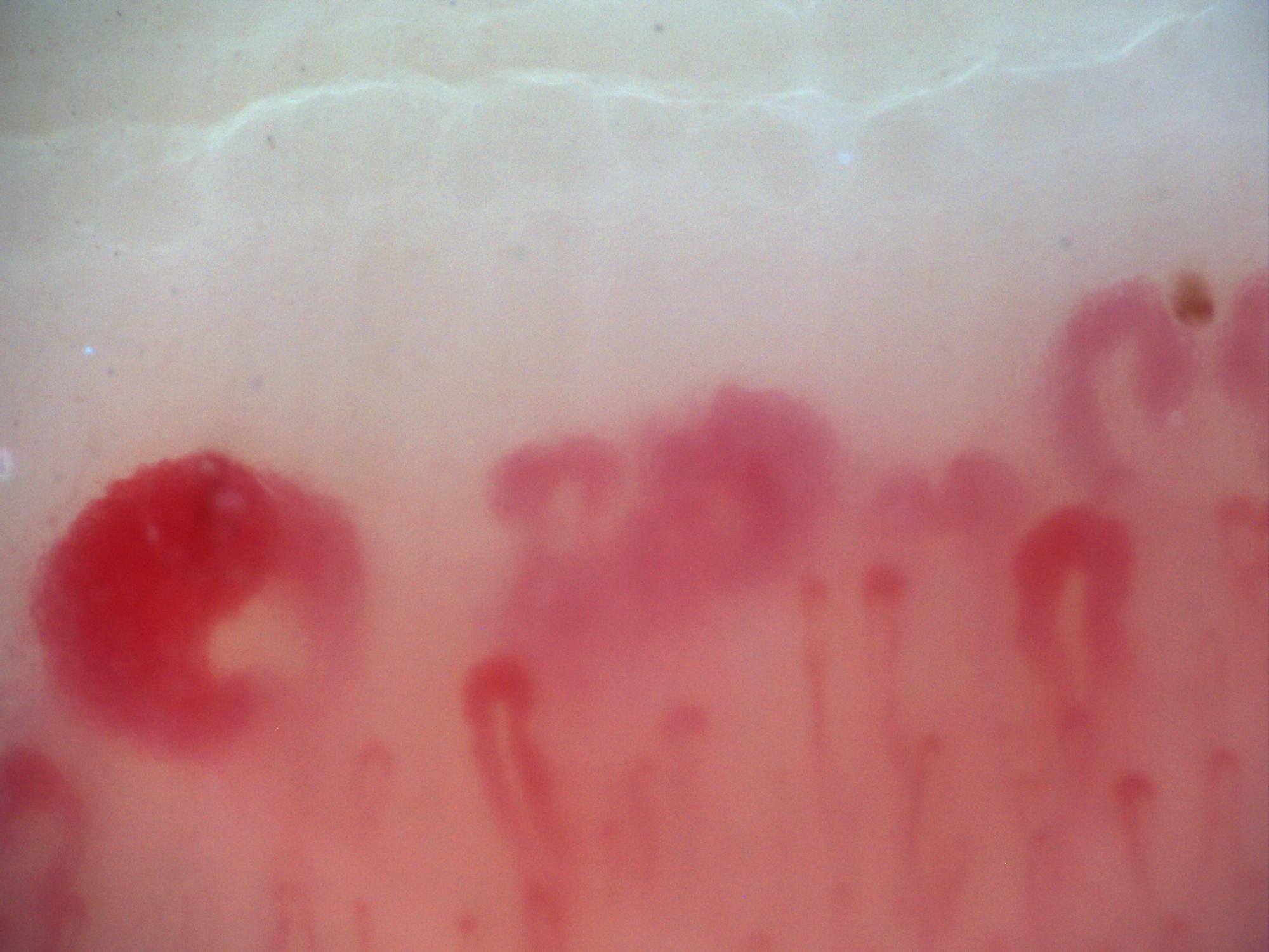
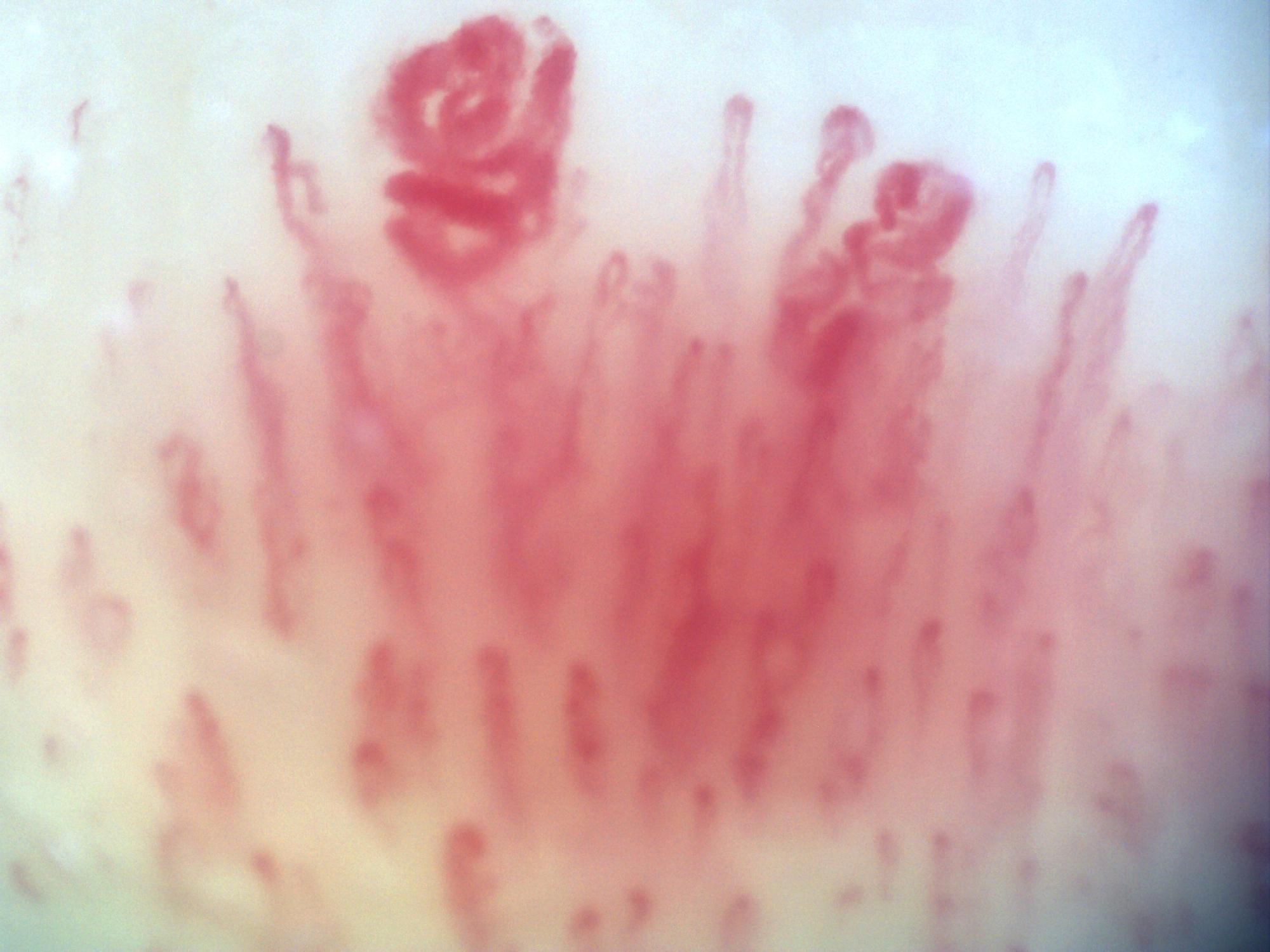
200x nailfold capillaroscopy field with prominent megacapillaries in an active scleroderma pattern
View details

Active scleroderma pattern with megacapillaries (field 2)

Author: Miguel Antonio Mesa Navas

Systemic sclerosisActive scleroderma patternModerate to Severe
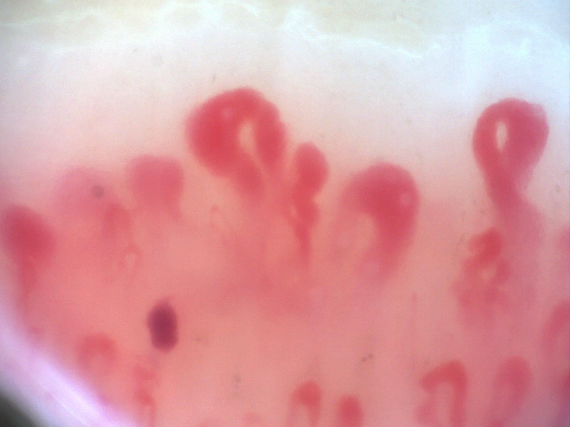
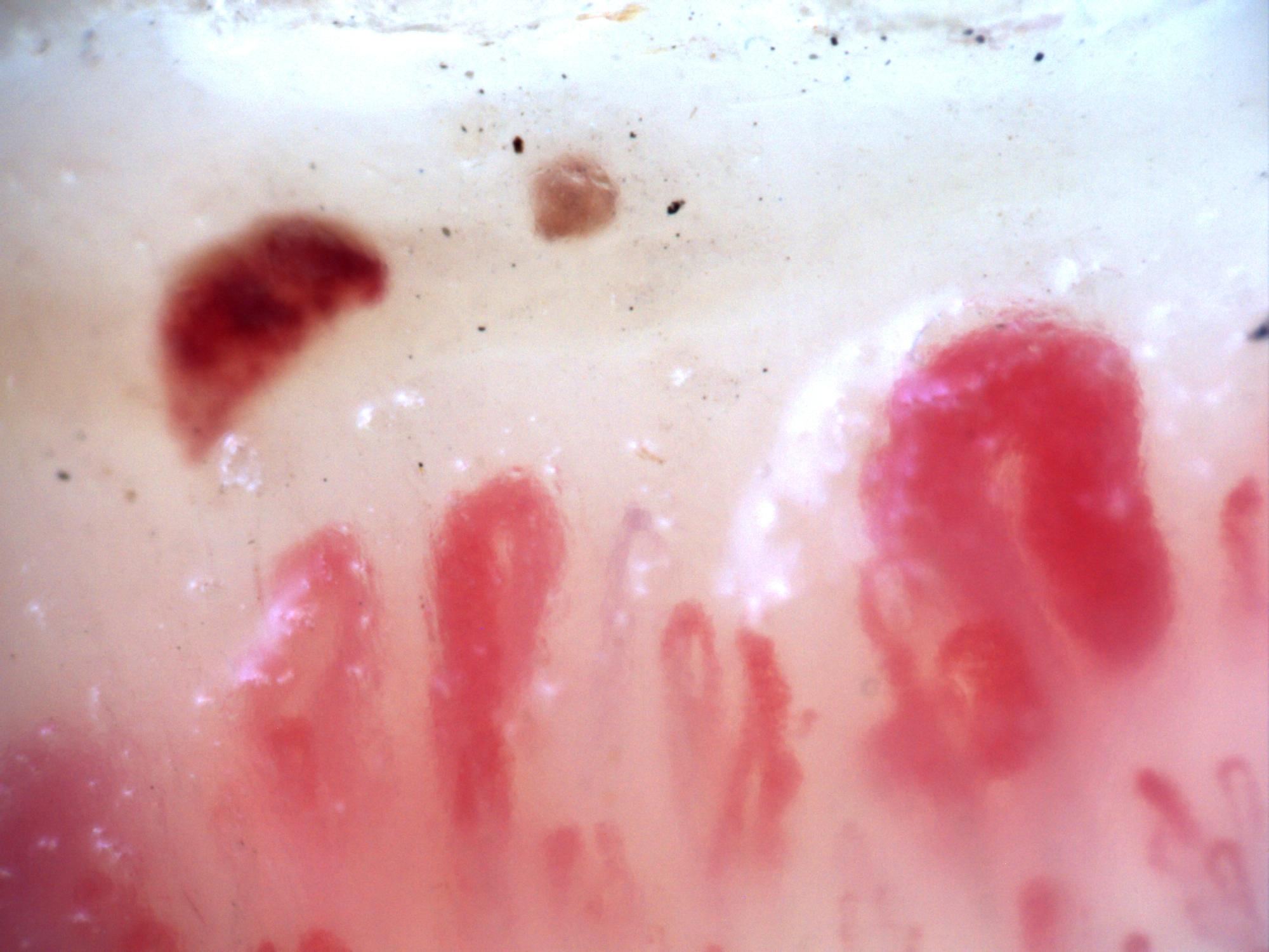
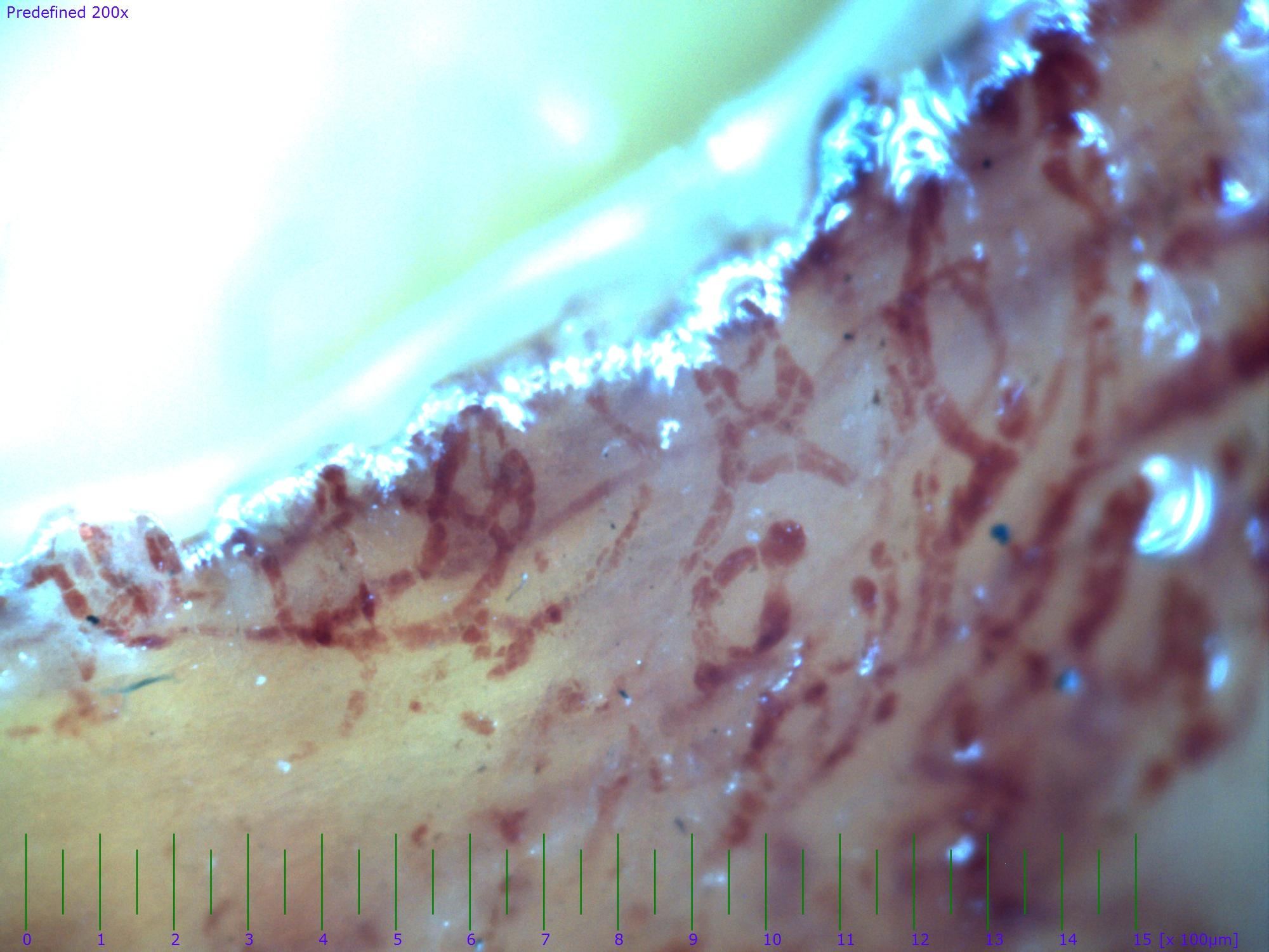
200x nailfold capillaroscopy field with visible megacapillaries in an active scleroderma pattern
View details

Active scleroderma pattern with megacapillaries (field 3)

Author: Miguel Antonio Mesa Navas

Systemic sclerosisActive scleroderma patternModerate to Severe
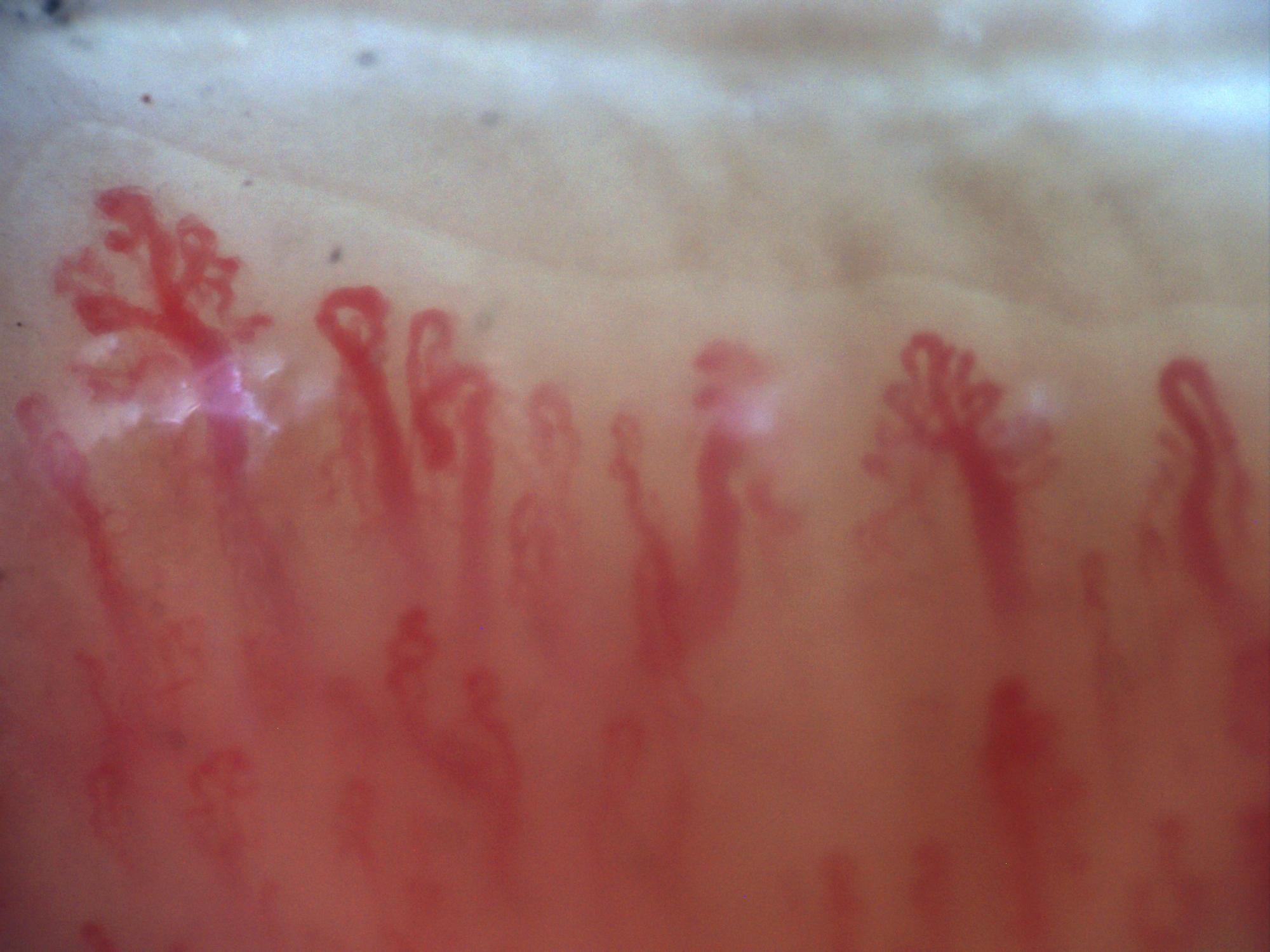
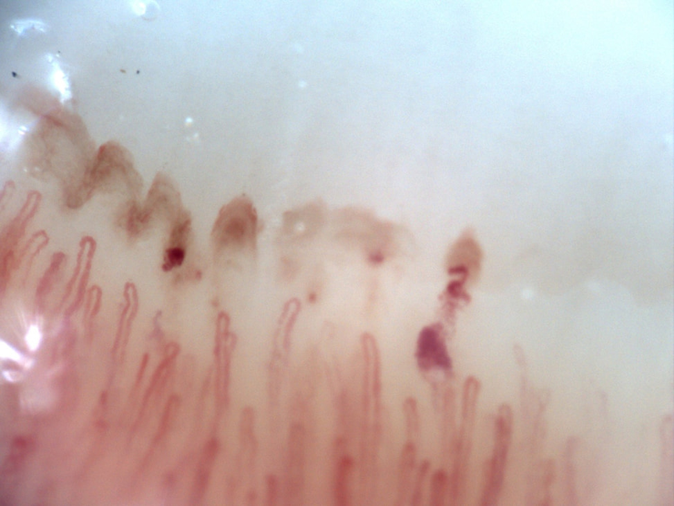
200x nailfold capillaroscopy field with large megacapillaries in an active scleroderma pattern
View details

Active scleroderma pattern with megacapillaries (field 4)

Author: Miguel Antonio Mesa Navas

Systemic sclerosisActive scleroderma patternModerate to Severe
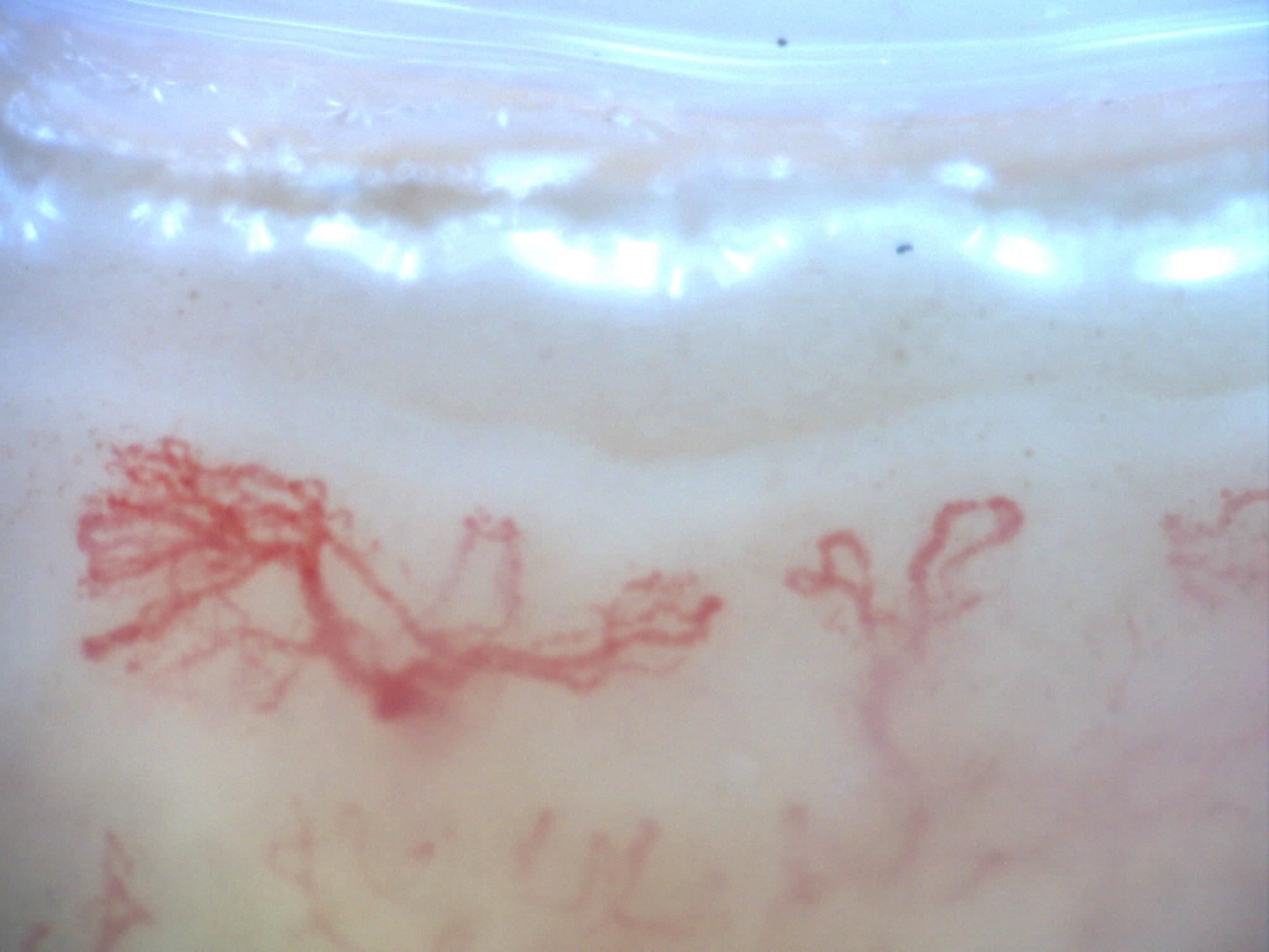
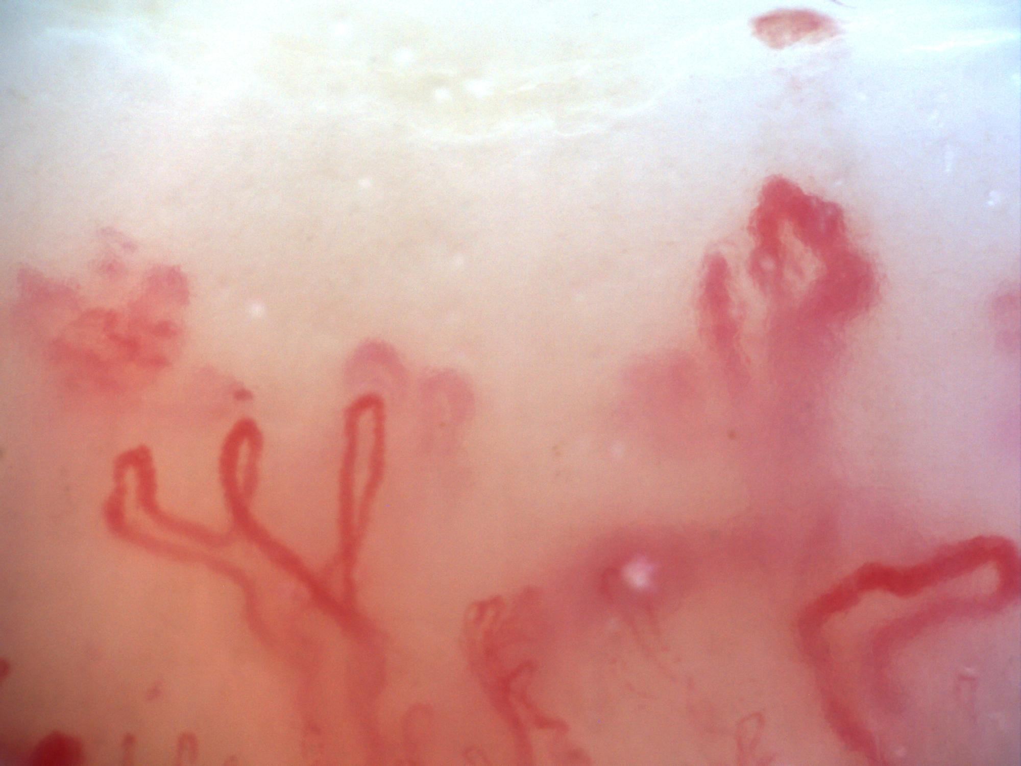
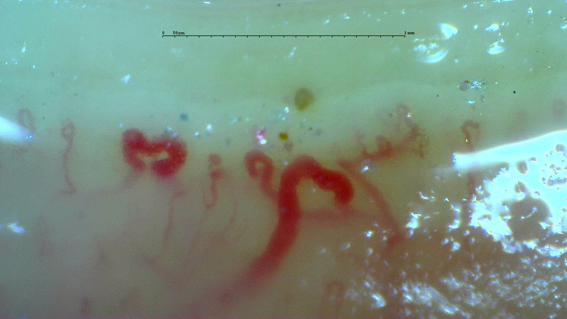
200x nailfold capillaroscopy field with a branching capillary on the left side
View details

Branching capillary on the left side

Author: Miguel Antonio Mesa Navas

UnspecifiedBranching capillary / neoangiogenesisUnspecified
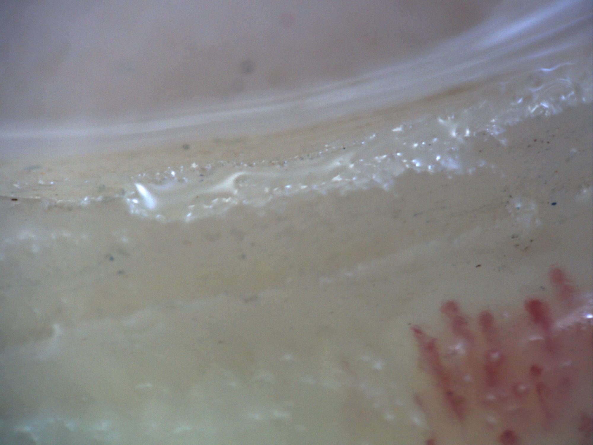
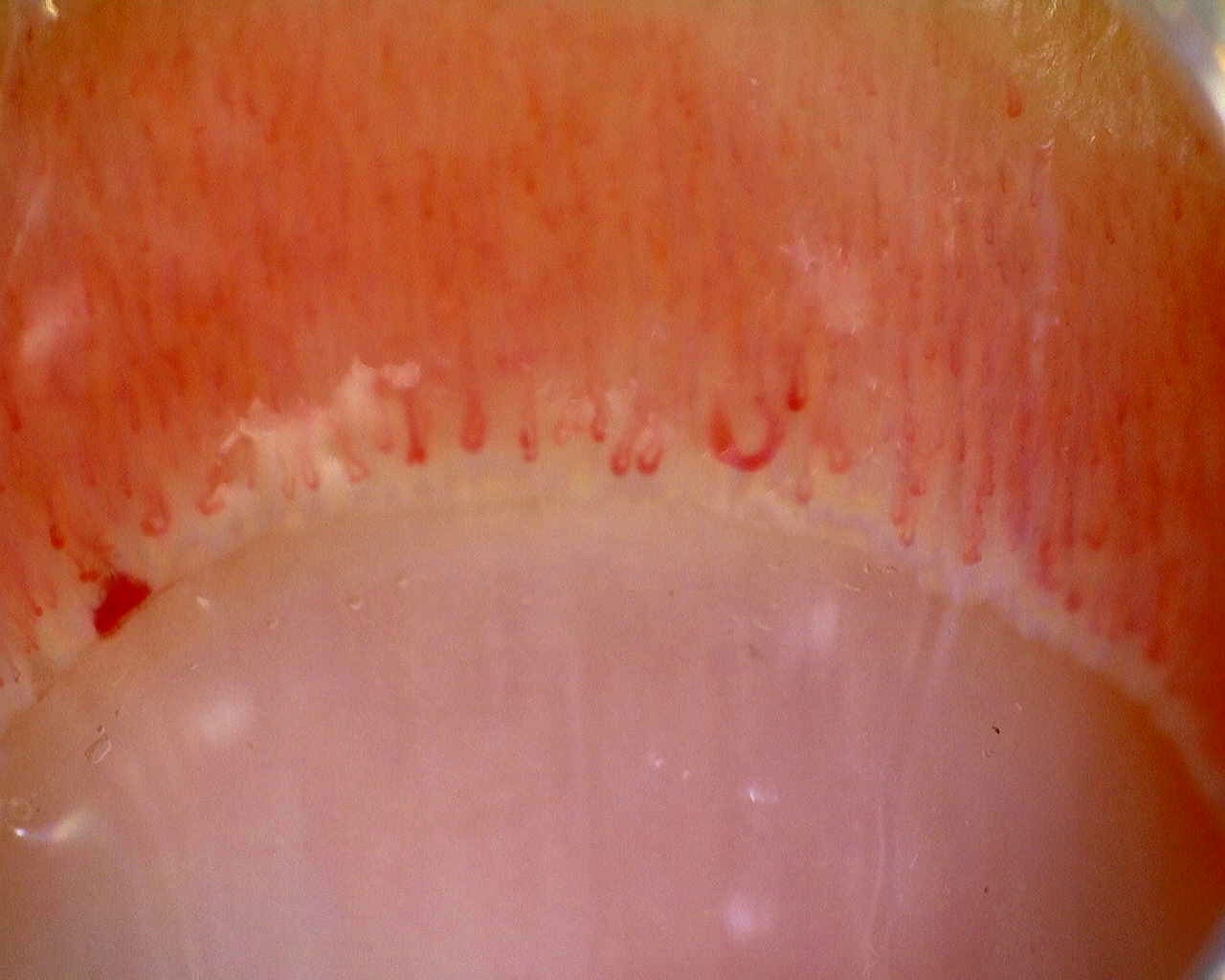
200x capillaroscopy field with disappearance of the capillary bed on the left margin due to excessive pressure on the nail
View details

Pressure artifact with loss of visible capillary bed

Author: Miguel Antonio Mesa Navas

Acquisition artifactPressure artifactUnspecified
200x capillaroscopy field showing a broad capillary-free space compatible with an avascular area
View details

Extensive avascular area

Author: Miguel Antonio Mesa Navas

UnspecifiedExtensive avascular areaSevere
200x capillaroscopy field from systemic sclerosis showing marked architectural disorganization and neoangiogenesis without megacapillaries
View details

Late scleroderma pattern with severe disorganization (field 1)

Author: Miguel Antonio Mesa Navas

Systemic sclerosisLate scleroderma patternSevere
200x capillaroscopy field from systemic sclerosis showing severe architectural disorganization and neoangiogenesis without megacapillaries
View details

Late scleroderma pattern with severe disorganization (field 2)

Author: Miguel Antonio Mesa Navas

Systemic sclerosisLate scleroderma patternSevere
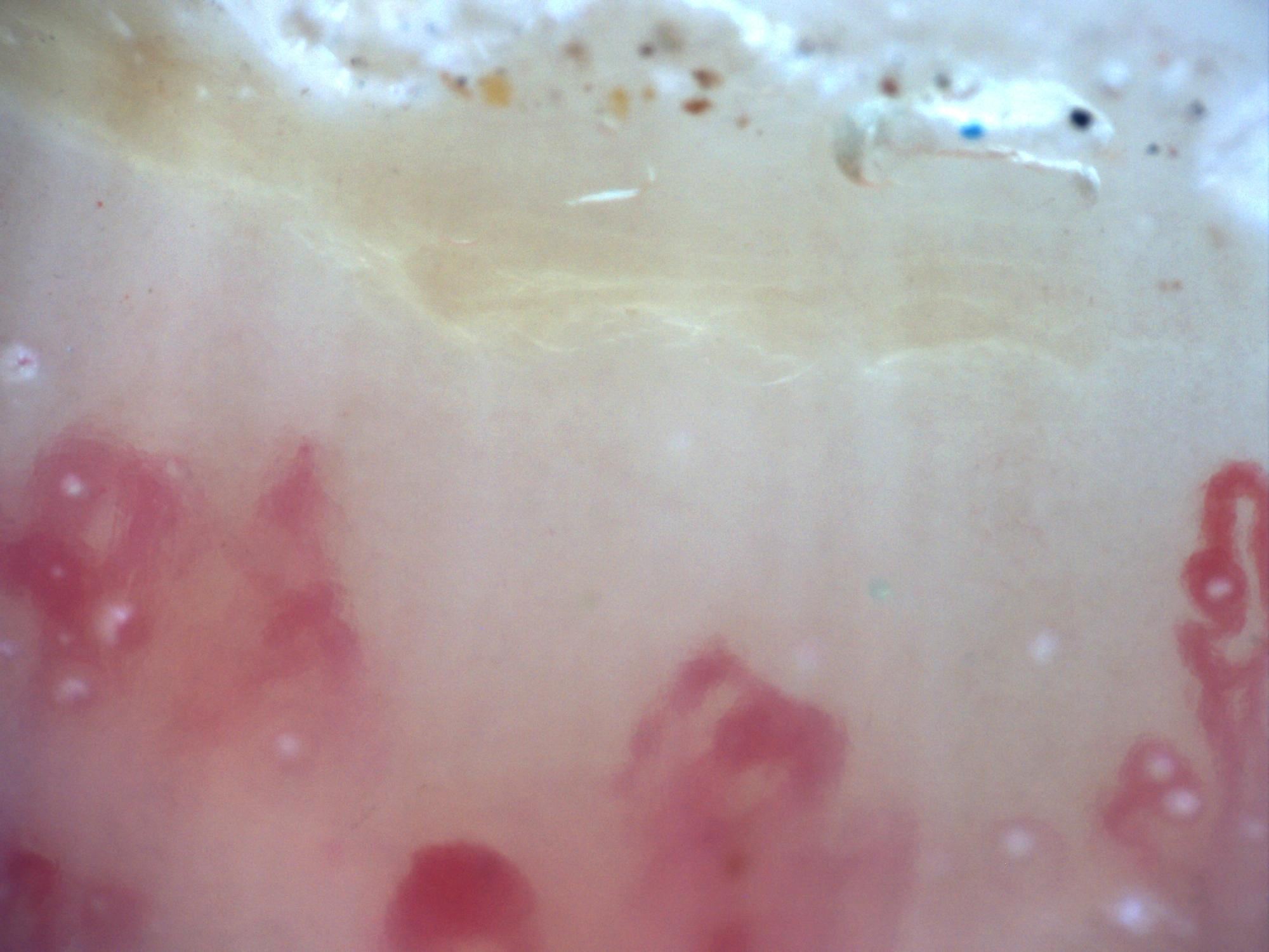
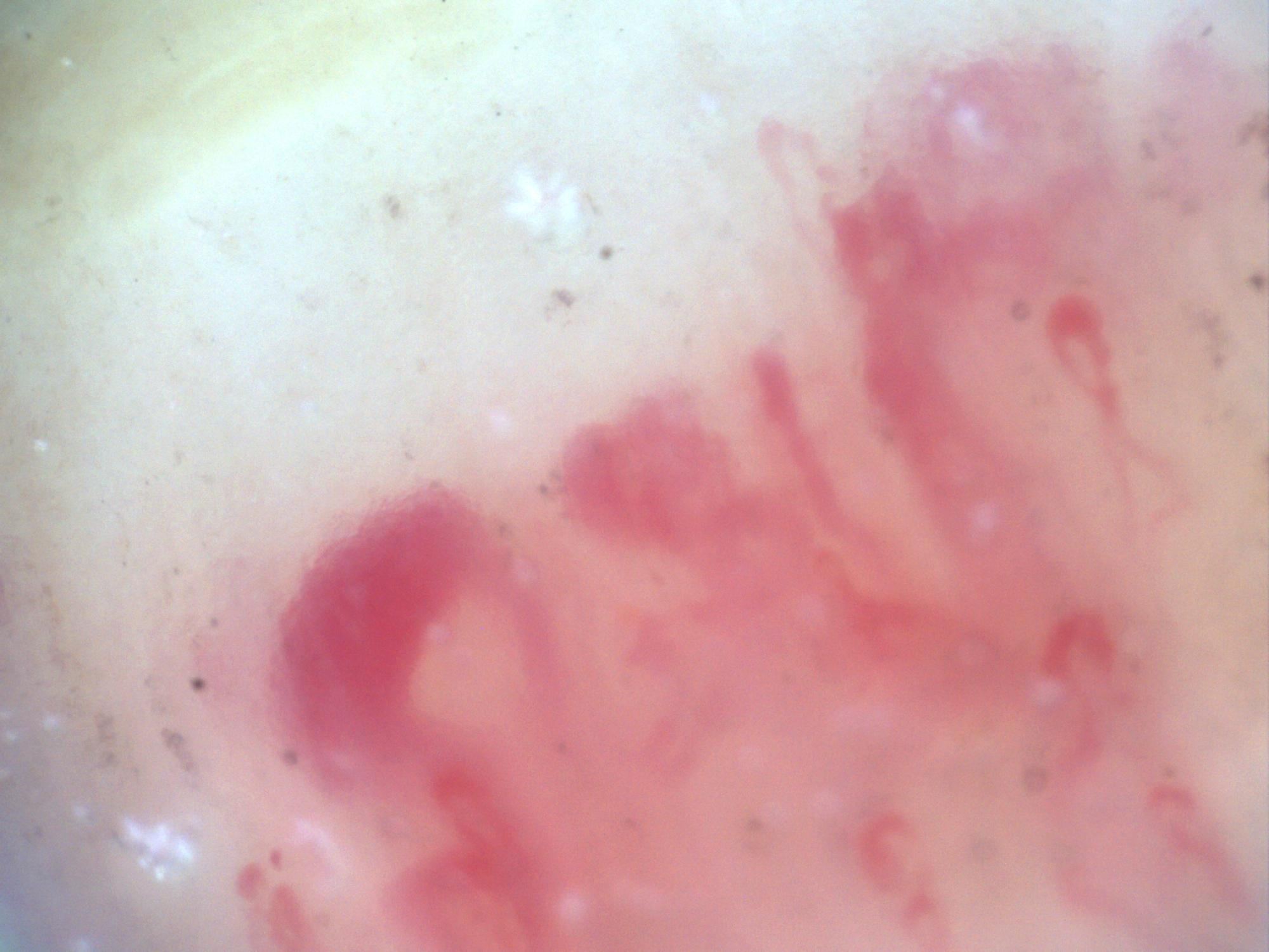
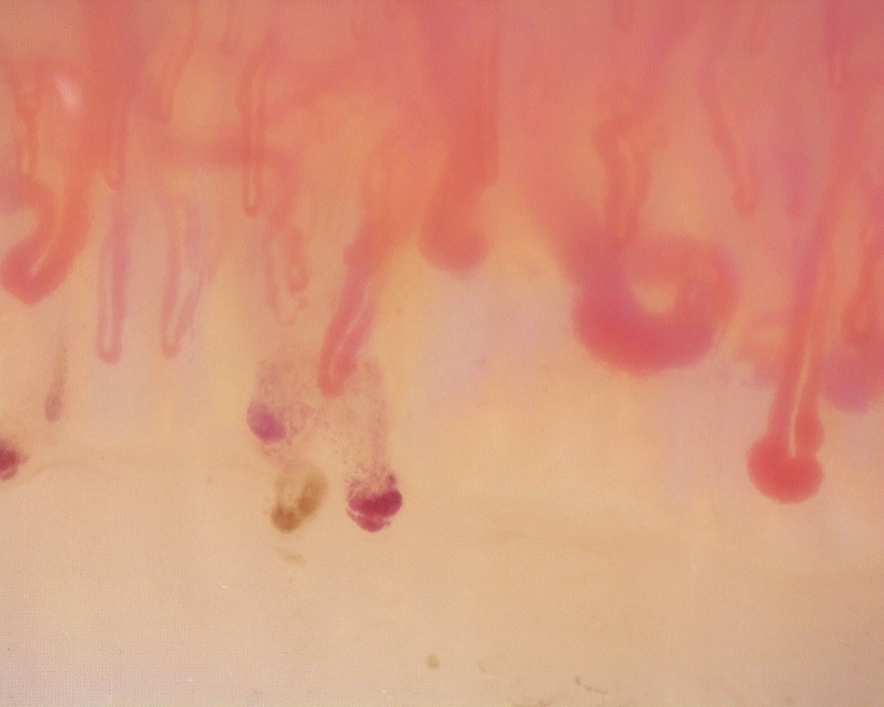
200x capillaroscopy field with a fog-like pericapillary haze between capillaries consistent with pericapillary edema
View details

Pericapillary edema on an active background

Author: Miguel Antonio Mesa Navas

Systemic sclerosisActive scleroderma patternModerate to Severe
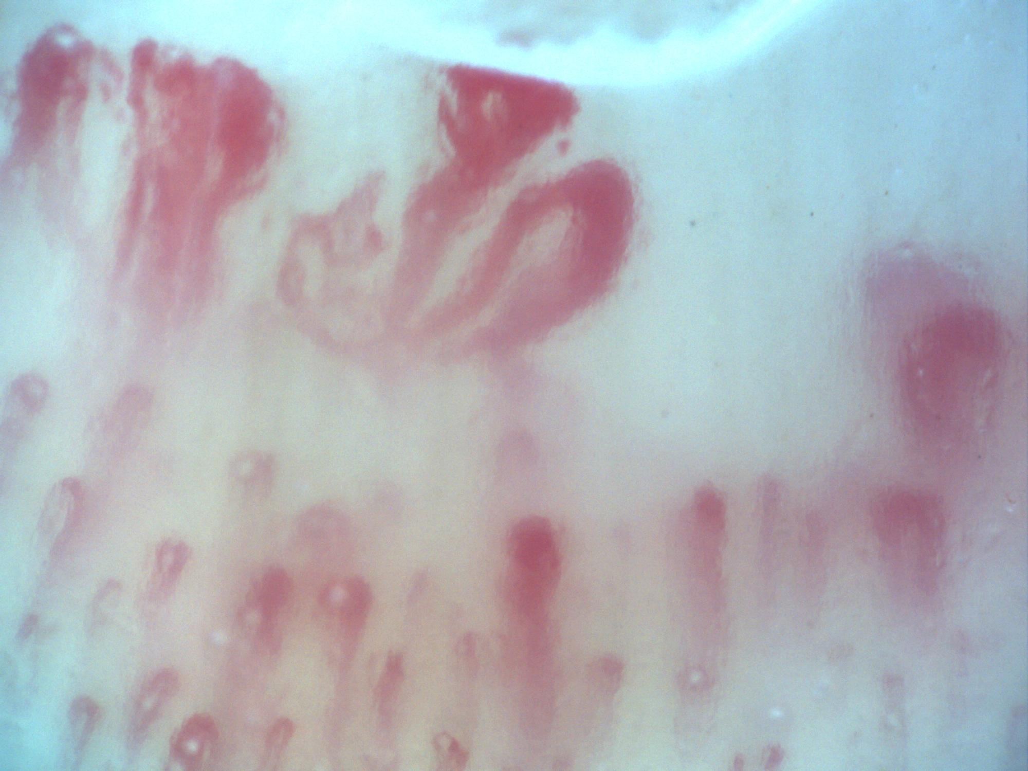
200x capillaroscopy field in systemic sclerosis showing megacapillaries with a ground-glass stromal appearance suggestive of fibrosis
View details

Ground-glass stromal fibrosis with megacapillaries (field 1)

Author: Miguel Antonio Mesa Navas

Systemic sclerosisActive scleroderma pattern with stromal fibrosisSevere
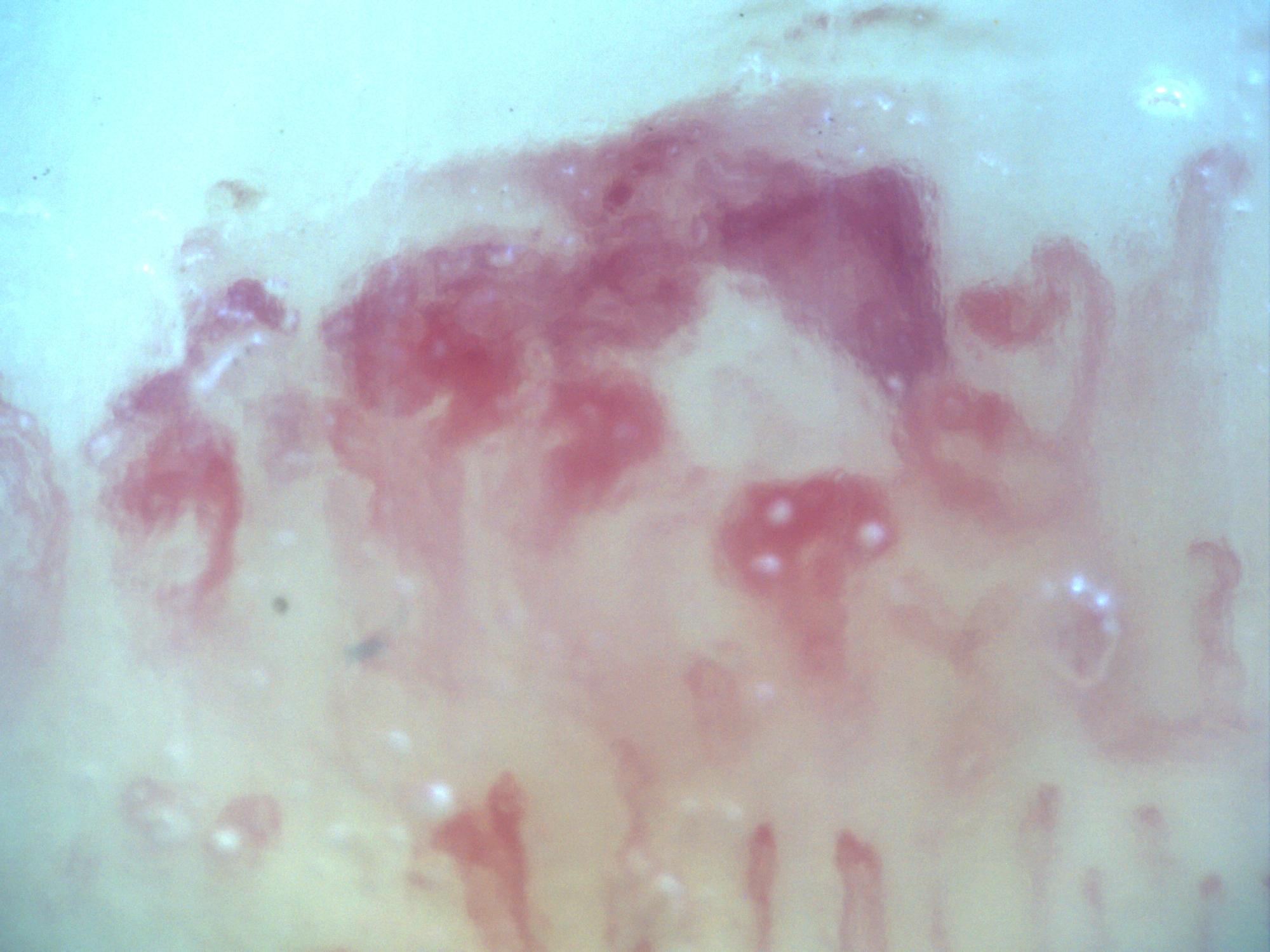
200x capillaroscopy field in systemic sclerosis showing megacapillaries, pericapillary edema, and a ground-glass stromal pattern
View details

Ground-glass stromal fibrosis with megacapillaries (field 2)

Author: Miguel Antonio Mesa Navas

Systemic sclerosisActive scleroderma pattern with stromal fibrosisSevere
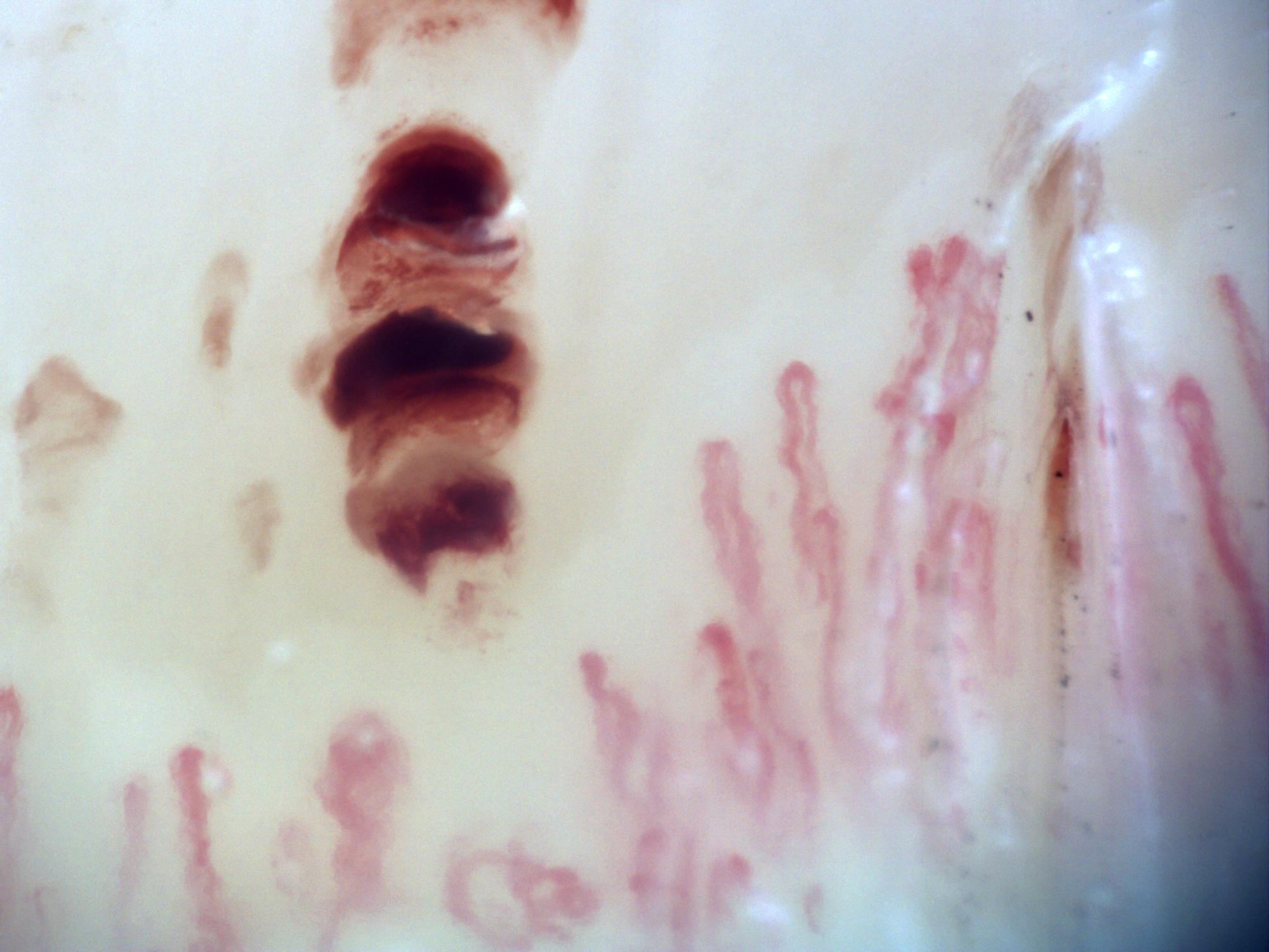
200x capillaroscopy field in systemic sclerosis showing a hemorrhage with a smoke-plume appearance
View details

Hemorrhage with smoke-plume appearance

Author: Miguel Antonio Mesa Navas

Systemic sclerosisHemorrhagic lesionModerate to Severe
200x capillaroscopy field in systemic sclerosis showing a representative microvascular hemorrhagic lesion
View details

Hemorrhagic field in systemic sclerosis

Author: Miguel Antonio Mesa Navas

Systemic sclerosisHemorrhagic lesionModerate to Severe
200x capillaroscopy field capturing a hemorrhage at the moment of its formation
View details

In situ hemorrhage formation

Author: Miguel Antonio Mesa Navas

Systemic sclerosisHemorrhagic lesionModerate to Severe
200x capillaroscopy field in systemic sclerosis showing a very large megacapillary
View details

Large megacapillary (field 1)

Author: Miguel Antonio Mesa Navas

Systemic sclerosisMegacapillary-dominant fieldModerate to Severe
200x capillaroscopy field in systemic sclerosis showing a prominent megacapillary
View details

Large megacapillary (field 2)

Author: Miguel Antonio Mesa Navas

Systemic sclerosisMegacapillary-dominant fieldModerate to Severe
200x capillaroscopy field in systemic sclerosis showing very dilated megacapillaries
View details

Large megacapillary (field 3)

Author: Miguel Antonio Mesa Navas

Systemic sclerosisMegacapillary-dominant fieldModerate to Severe
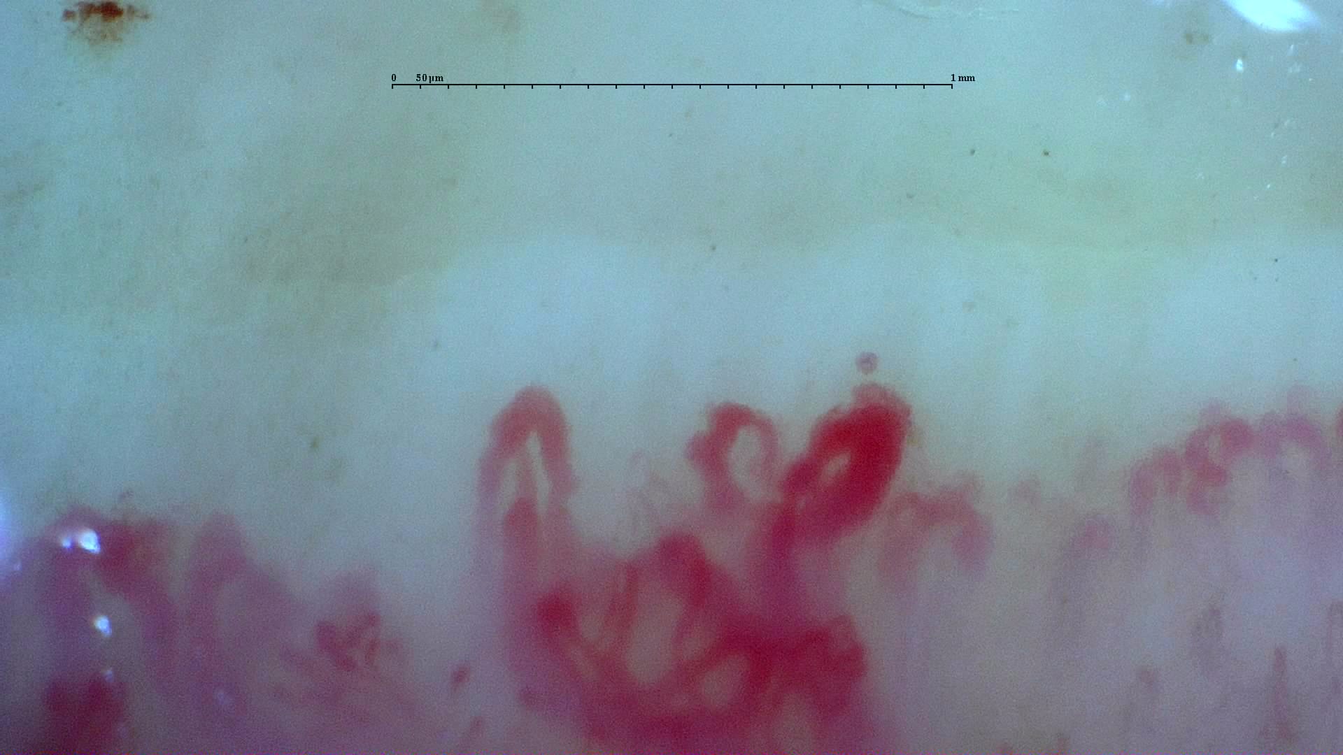
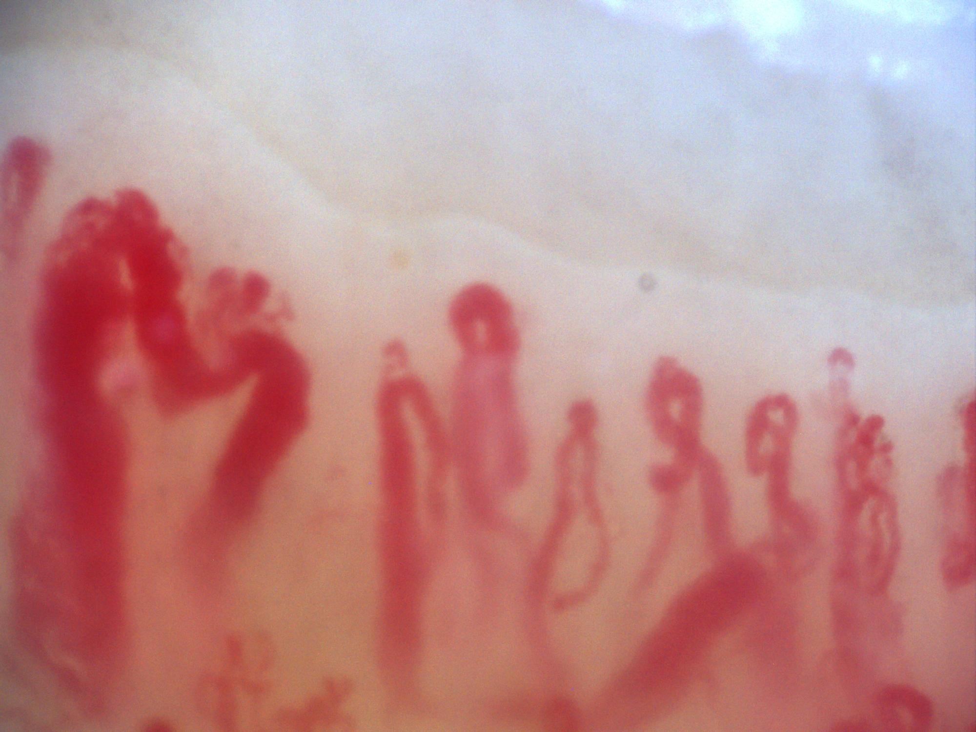
200x capillaroscopy field showing a giant capillary with a coiled appearance
View details

Coiled giant capillary

Author: Miguel Antonio Mesa Navas

Systemic sclerosisCoiled giant capillaryModerate to Severe
Capillaroscopy field from a critically ischemic digit with diffuse capillary thrombosis
View details

Suspected capillary thrombosis in critical digital ischemia

Author: Miguel Antonio Mesa Navas

Critical digital ischemiaSuspected capillary thrombosisSevere
Nailfold video capillaroscopy of the right fourth finger showing preserved capillary density with hemorrhages and no giant capillaries
View details

Preserved capillary density with anticoagulation-related hemorrhages

Author: Luis Peñaranda Bolaño

Anticoagulation-related hemorrhagesPreserved capillary density with hemorrhagesMild
Nailfold video capillaroscopy in anti-TIF-1gamma antibody-positive dermatomyositis showing enlarged, giant, and ramified capillaries
View details

Dermatomyositis with giant and ramified capillaries

Author: Luis Peñaranda Bolaño

DermatomyositisMyositis-associated microangiopathy with neoangiogenesisSevere
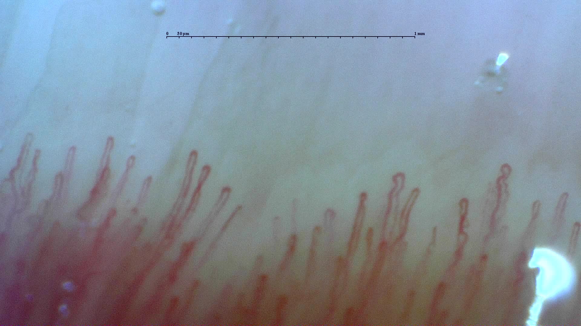
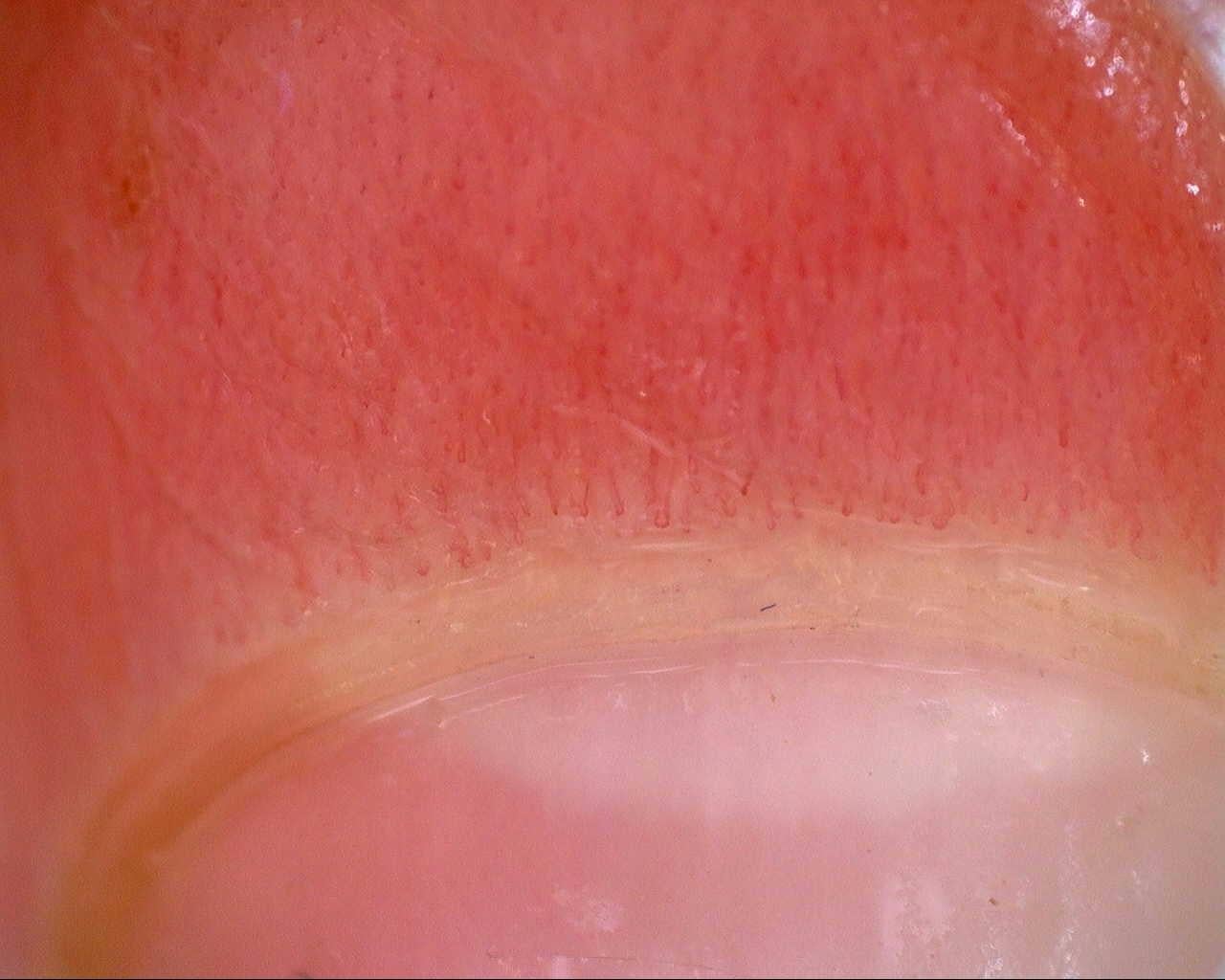
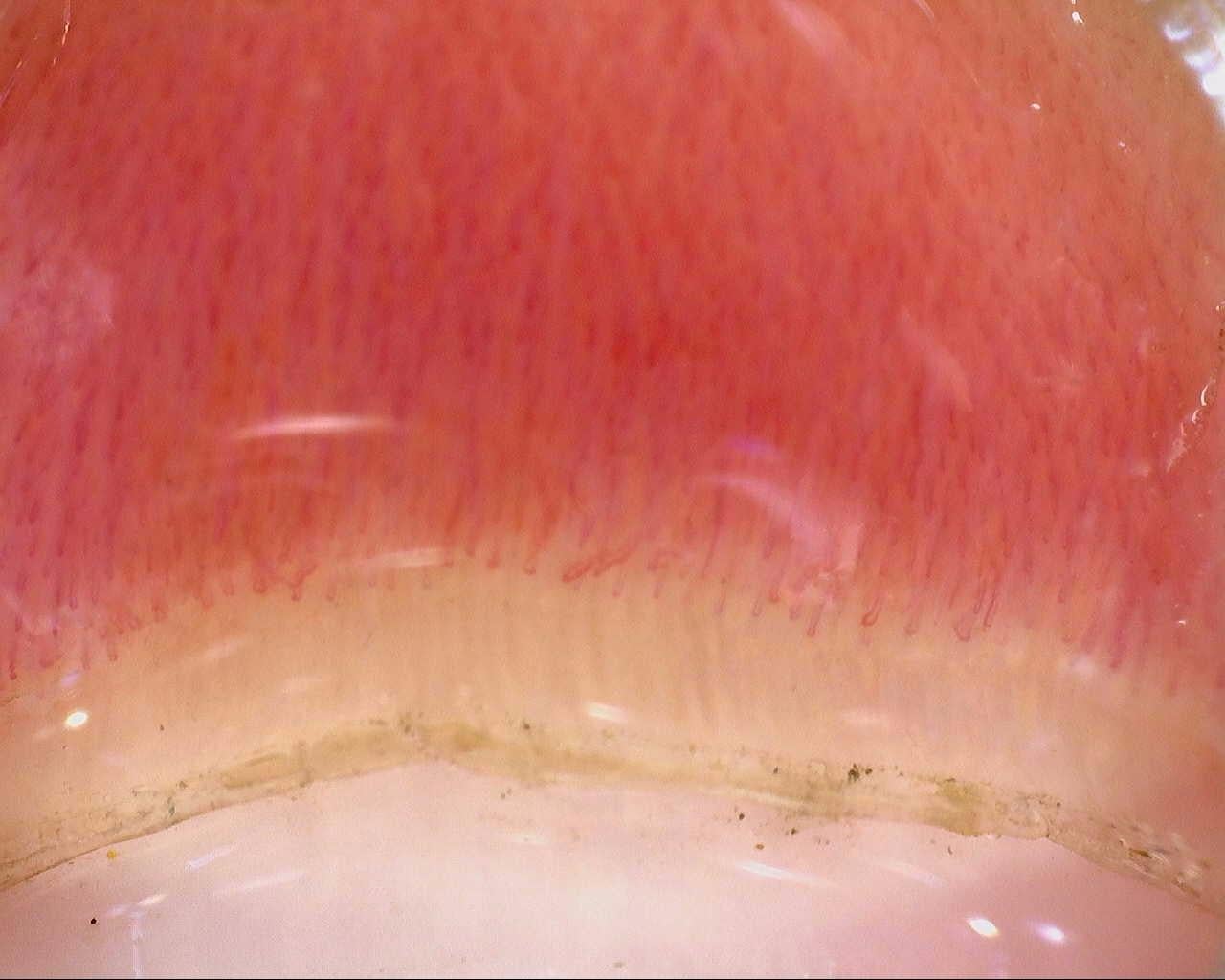
A field from the normal reference study, showing the distal row of narrow loops. Use the full study to compare density and shape across fingers.
View details

Normal reference: distal capillary row

Author: Miguel Antonio Mesa Navas

Normal CapillaroscopyNormal PatternSee complete study
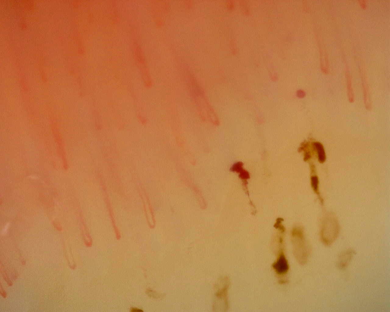
Dark hemorrhagic deposits alongside capillary loops in the nonspecific reference study. Hemorrhage alone does not define a scleroderma pattern.
View details

Nonspecific pattern: hemorrhagic field

Author: Miguel Martín Cascón

Non-Specific MicroangiopathyNon-Specific PatternSee complete study
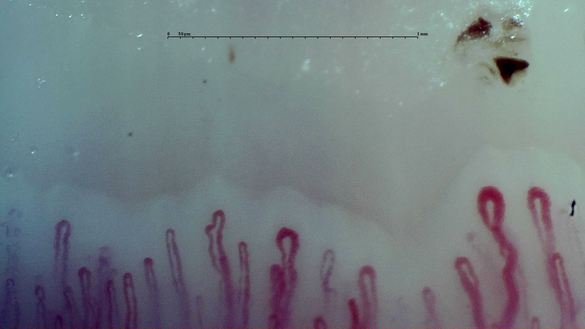
Selected field from the early-pattern reference study, with enlarged loops among a relatively preserved capillary row. The subtype belongs to the complete examination.
View details

Early scleroderma pattern: selected field

Author: Borja Gracia Tello

Systemic SclerosisEarly Cutolo SSc PatternSee complete study
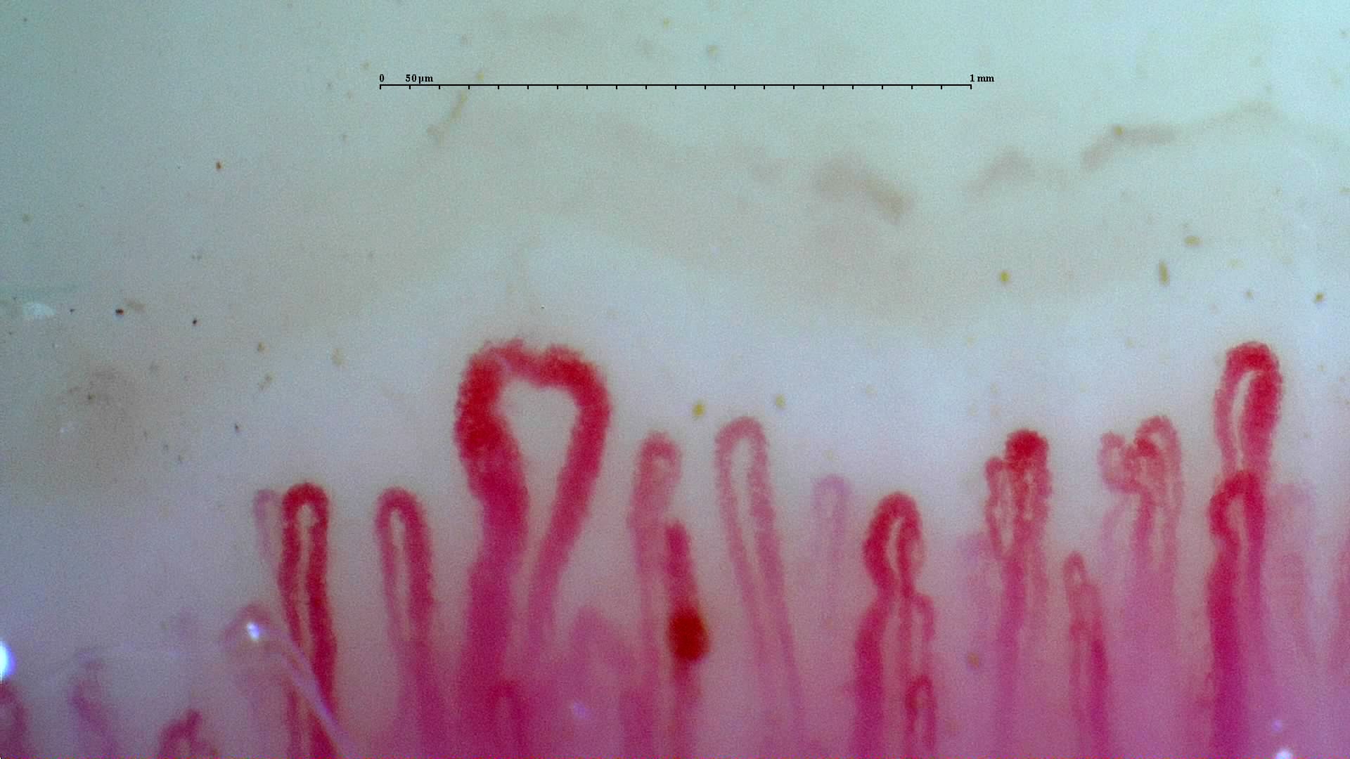
A prominent enlarged loop in the active-pattern reference study. Compare the neighboring capillaries and the other fields before assigning a pattern.
View details

Active scleroderma pattern: giant loop

Author: Borja Gracia Tello

Systemic SclerosisActive Cutolo SSc PatternSee complete study
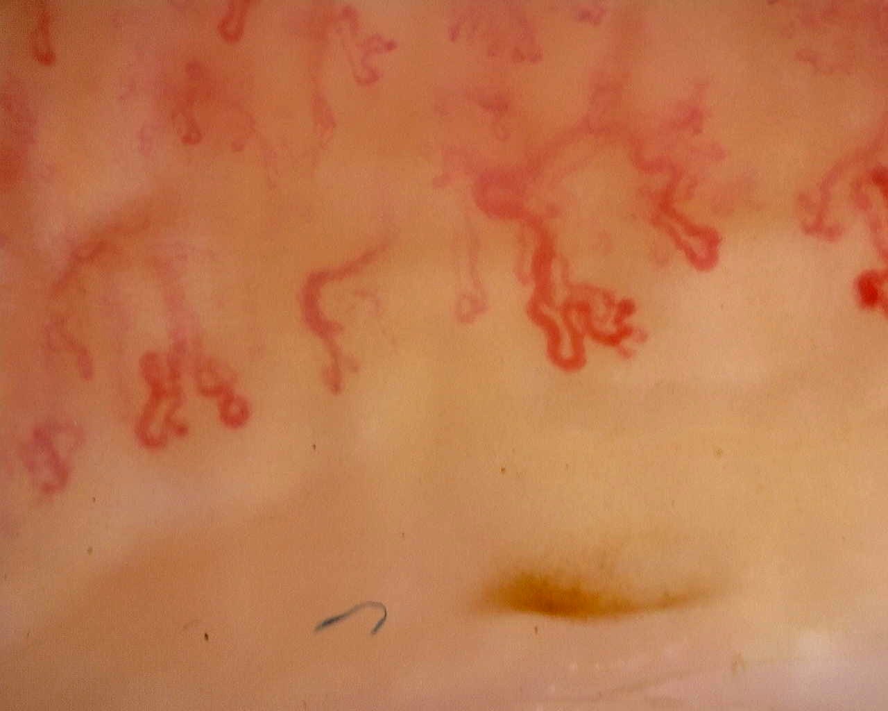
Irregular, ramified loops in a field from the late-pattern reference study. The full study provides context for capillary loss and distribution.
View details

Late scleroderma pattern: disorganized loops

Author: Gema María Lledó Ibáñez

Systemic SclerosisLate Cutolo SSc PatternSee complete study
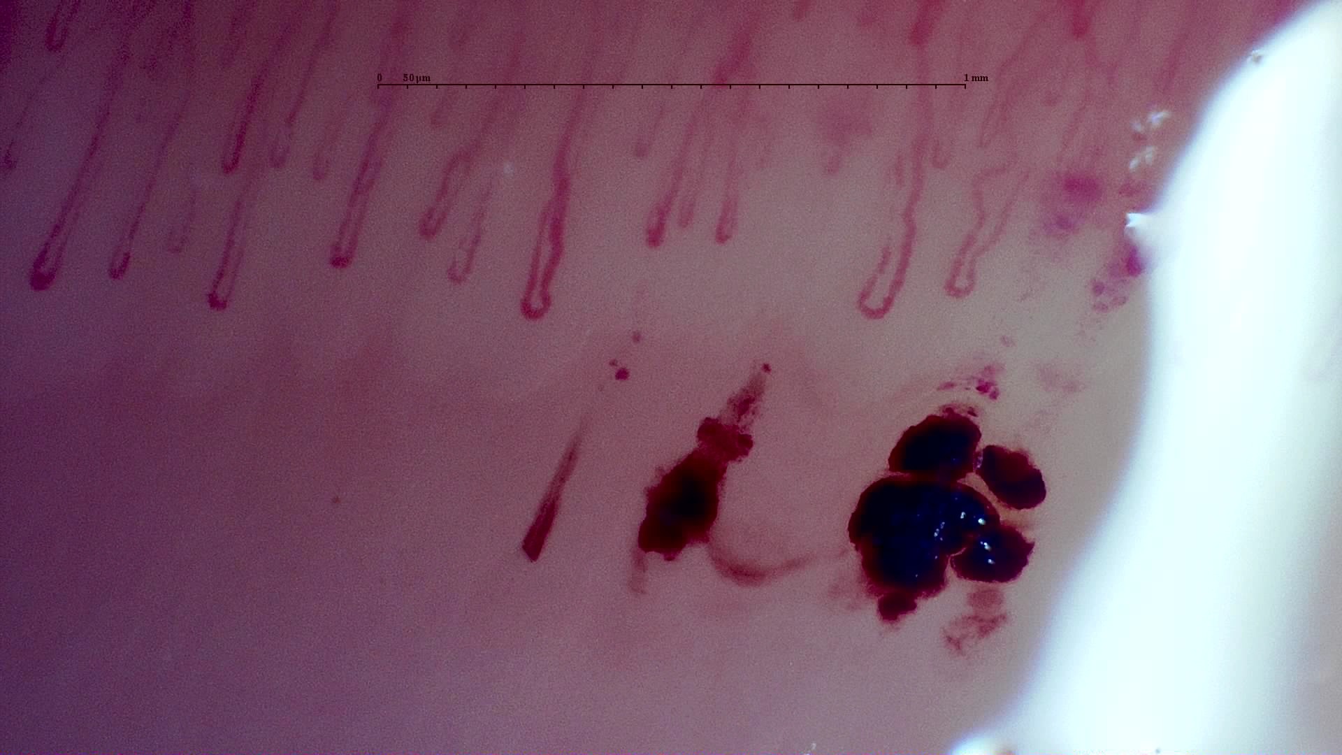
Large dark hemorrhagic deposits below the capillary row in the published MPO-ANCA vasculitis case. Its complete study is described as a nonspecific pattern without giants.
View details

MPO-ANCA vasculitis: hemorrhagic deposits

Author: Gema María Lledó Ibáñez

ANCA-Associated Vasculitis (MPO+)Non-Specific PatternSee complete study
An overview of the nailfold from the anti-Mi2β dermatomyositis case. This wider view provides context; use the complete study for calibrated measurements and individual capillary assessment.
View details

Anti-Mi2β dermatomyositis: overview

Author: Gema María Lledó Ibáñez

Dermatomyositis (anti-Mi2β+)Severe Myositis-Associated MicroangiopathySee complete study
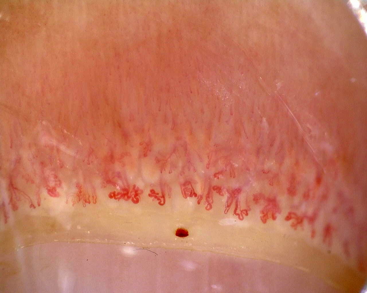
Overview of the nailfold in the ACA-positive active-pattern case. The reported density and giant-capillary percentage describe the study, not this photograph alone.
View details

ACA-positive systemic sclerosis: overview

Author: Gema María Lledó Ibáñez

Limited Cutaneous Systemic Sclerosis (ACA+)Active Cutolo Scleroderma PatternSee complete study
Enlarged loops and dark hemorrhagic deposits in the anti-Scl-70-positive active-pattern case. The source report notes no avascular areas in the study.
View details

Anti-Scl-70 systemic sclerosis: enlarged loops and hemorrhages

Author: Gela Gunashvili

Diffuse Systemic Sclerosis (anti-Scl-70+)Active Cutolo Scleroderma PatternSee complete study
Irregular enlarged loops in the anti-U3 RNP case. Although giants remain present, the final report classifies the overall microangiopathy as late.
View details

Anti-U3 RNP systemic sclerosis: irregular loops

Author: Miguel Antonio Mesa Navas

Systemic Sclerosis (anti-U3 RNP+)Late Scleroderma PatternSee complete study
A disorganized field with enlarged loops and surface reflections from the inter-digit heterogeneity case. Other fields differ markedly; inspect the full study and its image-quality caveats.
View details

Inter-digit heterogeneity: selected abnormal field

Author: Miguel Antonio Mesa Navas

Systemic Sclerosis (Anticentromere-Positive)Heterogeneous Scleroderma Pattern (Active to Late)See complete study
Irregular and branching forms in the anti-RNP-positive mixed connective tissue disease case. The source report describes a scleroderma-like pattern without an assignable Cutolo subtype.
View details

Mixed connective tissue disease: irregular branching forms

Author: Miguel Antonio Mesa Navas

Mixed Connective Tissue Disease (anti-RNP+)Scleroderma-Like Pattern (Unclassifiable Cutolo Subtype)See complete study
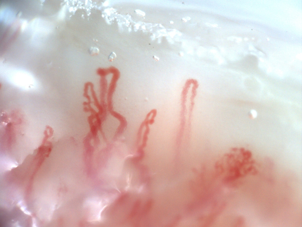
Prominent enlarged loops and a complex branching form in the initial anti-NXP2 dermatomyositis study. Compare this with the linked follow-up image and complete serial studies.
View details

Anti-NXP2 dermatomyositis: initial enlarged loops

Author: Luis Saez Comet

Dermatomyositis (anti-NXP2+)Myositis-Associated Microangiopathy with Treatment ResponseSee complete study
Follow-up field from the adult anti-NXP2 dermatomyositis case. The source report documents improved study-wide density and disappearance of giants and hemorrhages; these are longitudinal study findings.
View details

Anti-NXP2 dermatomyositis: follow-up field

Author: Luis Saez Comet

Dermatomyositis (anti-NXP2+)Follow-up studySee complete study
Complex, winding and ramified vascular forms in the anti-TIF1γ dermatomyositis case. A scleroderma-like image appearance does not establish systemic sclerosis.
View details

Anti-TIF1γ dermatomyositis: complex vascular shapes

Author: Franklin Uguña Sari

Dermatomyositis (anti-TIF1γ+)Scleroderma-Like PatternSee complete study
Nailfold overview from the juvenile dermatomyositis case with Gottron papules. The complete report describes the vascular abnormalities; this image is an orientation view.
View details

Juvenile dermatomyositis: nailfold overview

Author: Clara Udaondo

Juvenile DermatomyositisLate Scleroderma-Like PatternSee complete study
Initial nailfold overview in the juvenile anti-NXP2 longitudinal case. Compare the follow-up and the documented quantitative findings in the complete case.
View details

Juvenile anti-NXP2 dermatomyositis: initial overview

Author: Clara Udaondo

Juvenile Dermatomyositis (anti-NXP2+)Early Scleroderma-Like Pattern with Treatment ResponseSee complete study
Follow-up nailfold overview from the same juvenile anti-NXP2 case. Differences in overview appearance should be interpreted alongside acquisition conditions and the serial reports.
View details

Juvenile anti-NXP2 dermatomyositis: follow-up overview

Author: Clara Udaondo

Juvenile Dermatomyositis (anti-NXP2+)Follow-up studySee complete study
Overview from the active juvenile anti-TIF1 dermatomyositis case. Open the source study to examine the arborizing capillaries described in its report.
View details

Juvenile anti-TIF1 dermatomyositis: nailfold overview

Author: Clara Udaondo

Juvenile Dermatomyositis (anti-TIF1+)Active Scleroderma-Like PatternSee complete study
Initial nailfold overview from the longitudinal mixed connective tissue disease case. Use the serial studies to assess change in morphology and density.
View details

Mixed connective tissue disease: initial overview

Author: Clara Udaondo

Mixed Connective Tissue Disease (anti-RNP+)Early Scleroderma Pattern with ImprovementSee complete study
Follow-up overview from the same mixed connective tissue disease case. The linked report places this image in the clinical and longitudinal context.
View details

Mixed connective tissue disease: follow-up overview

Author: Clara Udaondo

Mixed Connective Tissue Disease (anti-RNP+)Follow-up studySee complete study

Image Usage Guidelines

All images in this library are provided for educational purposes only. Images may be used for teaching and learning activities within medical institutions.

For publication or commercial use, please contact us for licensing information. Proper attribution is required for all uses.

Images have been anonymized and provided with appropriate consent for educational use.