Capillaroscopy in children and adolescents is not simply a smaller version of the adult examination. Test tolerance, some normal features, and above all the clinical weight of findings differ within a spectrum of less prevalent diseases with different trajectories. Current evidence mainly supports its use in pediatric Raynaud's phenomenon, juvenile systemic sclerosis (jSSc), juvenile dermatomyositis (JDM), and some overlap connective tissue diseases. In other conditions, it remains a supporting technique rather than a decisive criterion [1, 2, 3, 4, 5, 6, 7, 8, 9, 10, 11, 12, 13, 14].
- Pediatric normality does not exactly reproduce adult normality: healthy children may have more tortuous loops or mild morphological variants without microangiopathy [2, 3].
- In an international multicenter series of pediatric patients with rheumatic and musculoskeletal diseases, the scleroderma pattern occurred mainly in jSSc, JDM, and connective tissue diseases. Patterns in juvenile idiopathic arthritis (JIA) and primary Raynaud's phenomenon were much closer to those in healthy controls [2].
- In pediatric Raynaud's phenomenon, positive antinuclear antibodies (ANA) and capillary loss may inform prognosis, but capillaroscopy alone cannot reliably identify which child will develop a defined connective tissue disease [4, 5, 6, 7].
- The most consistent signal beyond jSSc occurs in JDM, where capillary loss, rarefaction, and diameter changes have been associated with activity and vascular damage [10, 11].
- In pediatric lupus and other childhood rheumatic diseases, capillaroscopy may detect abnormalities, but specificity remains limited and interpretation should be cautious and longitudinal [2, 12, 14].
- A good pediatric examination depends heavily on the setting: acclimatization, cooperation, finger size, avoiding excessive pressure, and recognizing that fewer high-quality images may be more informative than a complete but poorly acquired examination [1, 2].
Learning path
If you care for children or adolescents with Raynaud's phenomenon or suspected rheumatic disease, this unit explains what differs from adults and which findings require greater caution. It is especially useful if you are already familiar with normality and variability, Raynaud's phenomenon, and dermatomyositis and inflammatory myopathies [1, 2, 4, 5, 6, 7, 8, 9, 10, 11].
What changes in pediatrics
The first difference is practical: cooperation, finger size, anxiety, and acclimatization time have a greater effect on examination quality than in adults. The second is conceptual: the disease spectrum differs, and the pretest prevalence of systemic sclerosis (SSc) or defined connective tissue diseases is much lower [1, 2, 4].
A recent review focused on everyday clinical practice describes the setting well: pediatric capillaroscopy is useful, but requires knowledge of age-related normality, standardized interpretation, and follow-up recommendations that differ from those for adults [1]. This is particularly relevant in childhood Raynaud's phenomenon, where an abnormal finding may justify closer surveillance but does not carry the same classification weight as in an adult clinic with a high volume of connective tissue disease [1, 4, 5, 6, 7].
An international series published in 2023 provides a second practical message. Among 250 children assessed at 13 centers, the JIA and primary Raynaud's groups had qualitative findings very similar to healthy controls, while the clearest differences occurred in jSSc, JDM, and other connective tissue diseases [2]. These observations help set realistic expectations and avoid indiscriminate testing for any acral pain or mild vasomotor symptom.
Normality and variability
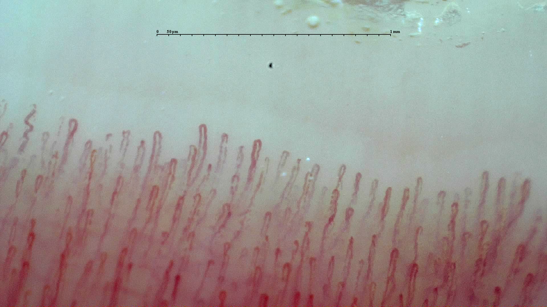
Understanding pediatric normality is essential because mild tortuosity or atypical morphology is relatively common in healthy children and may be overdiagnosed if adult frameworks are applied without adjustment [2, 3].
A reference study by Terreri et al in healthy children showed that most capillaroscopies were normal but not flawless: up to one-third could show subjectively tortuous patterns or minor changes without pathological significance [3]. Years later, the international cohort of pediatric patients and healthy children confirmed a similar picture: mean density in healthy controls was around 7.4 capillaries/mm. Compared with these controls, density was significantly lower in jSSc, JDM, mixed connective tissue disease, childhood-onset lupus, and localized scleroderma, but no relevant qualitative differences were found in JIA or primary Raynaud's phenomenon [2]. A pediatric examination should therefore not be classified as pathological because of a few tortuous loops or isolated shape variants.
The key is to look for coherent, reproducible patterns. In a healthy child, distal architecture is usually orderly, overall density is adequate, and there is no repeated combination of giant capillaries, capillary loss, and hemorrhages associated with structural damage. When these domains converge, the examination becomes much more informative and the findings go beyond simple variability [2, 3].
| Finding | In healthy children | When it is more concerning |
|---|---|---|
| Mild tortuosity | Relatively frequent [2, 3] | When accompanied by capillary loss, giant capillaries, or repeated hemorrhages |
| Capillary density | Around 7–8/mm [2] | When rarefaction is clear and reproducible across several fingers |
| Giant capillaries | Not expected as a dominant finding | When repeated or combined with hemorrhages [2, 8, 9, 10] |
| Disorganized architecture | Not a feature of normality | Should always be assessed in the context of possible microangiopathy |
Pediatric Raynaud's phenomenon and follow-up
Raynaud's phenomenon remains the main reason for pediatric capillaroscopy. The technique has real value here, chiefly for risk stratification and follow-up decisions rather than definitive diagnosis [1, 4, 5, 6, 7].
The 2016 European recommendations proposed that every child with Raynaud's phenomenon be assessed with ANA, SSc-specific antibodies, and capillaroscopy, using these three components to determine follow-up intensity [4]. This reasoning remains relevant: clearly abnormal capillaroscopy or positive ANA supports closer monitoring, while a stable normal pattern in a child without autoimmunity is more reassuring than in an adult [1, 4].
Predictive ability nevertheless remains imperfect. In a prospective cohort of 250 children with Raynaud's phenomenon, only 23.6% developed a defined rheumatic disease over one to six years of follow-up. Capillaroscopic abnormalities and ANA positivity were more frequent in this group, but no single rule provided accurate individual prediction [5]. More recently, another study found that capillary loss was associated with connective tissue disease development only in the ANA-negative subgroup, while adding capillaroscopy to ANA did not clearly improve overall prediction [6].
The relationship between capillaroscopic findings and autoantibodies also appears to differ between children and adults. A 2023 international analysis found that ANA were associated with dilated capillaries and reduced density in adults, but not in the pediatric cohort. This reinforces the need for age-specific interpretation [7].
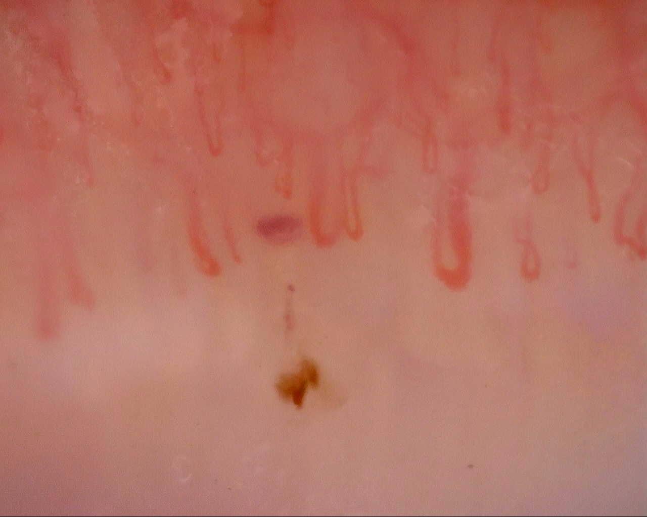 A. Initial examination · left ring finger · approximately 215×
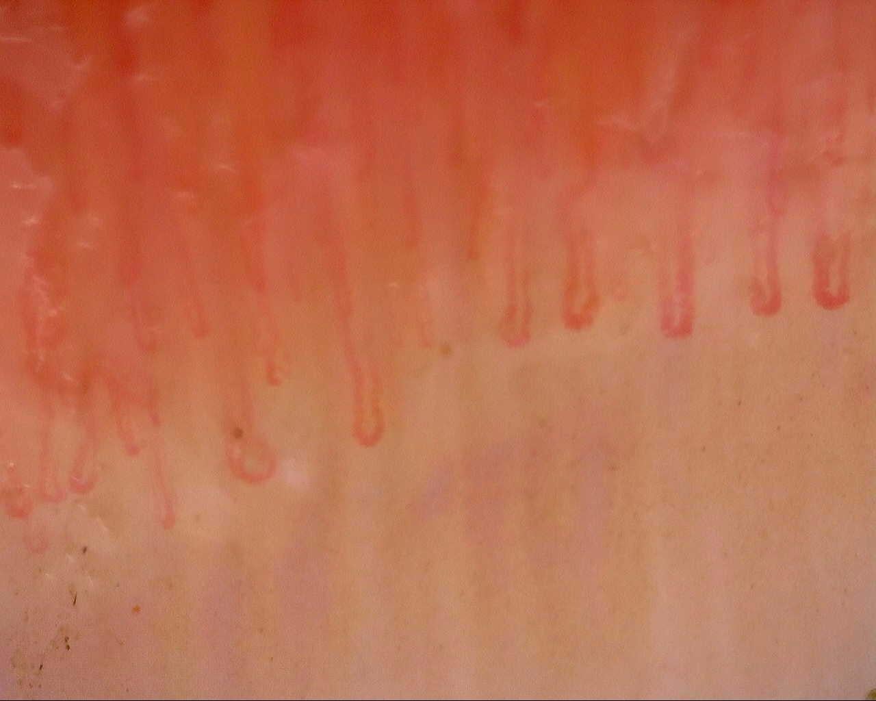 B. Follow-up · left ring finger · approximately 211×
Diseases in which capillaroscopy adds the most
Capillaroscopy does not have uniform value in pediatrics. It adds most in jSSc and JDM; in pediatric lupus, localized scleroderma, and other conditions, its role is more complementary [1, 2, 8, 9, 10, 11, 12, 13, 14].
Juvenile systemic sclerosis
The 2021 SHARE recommendations explicitly include capillaroscopy in baseline assessment and follow-up of jSSc [8]. This is consistent with earlier comparative literature: an exploratory EUSTAR study in patients who had reached adulthood found a scleroderma pattern in 93.3% of juvenile-onset cases versus 88% of adult-onset cases, with similar subpattern distributions [9]. In a child with Raynaud's phenomenon, swollen hands, or sclerodactyly, capillaroscopy carries substantial weight and may be among the most useful findings at the first assessment [8, 9].
Juvenile dermatomyositis
JDM is the other major pediatric indication. A 2023 study in untreated patients found lower density, greater tortuosity, and correlations between nailfold vasculopathy and clinical activity [10]. A 2025 study confirmed these observations: patients with active disease had lower total density and larger capillary diameters than those with inactive disease, supporting capillaroscopy as a complementary biomarker of vascular activity [11]. A systematic search of morphometric studies also suggests differences according to myositis-specific autoantibodies, although this aspect remains more exploratory [11, 14].
McClellan et al studied 76 participants, including 33 with JDM and eight with overlap myositis. Abnormal shapes, specifically capillaries classified as branched or ramified by automated analysis with Capillary.io, were more frequent in juvenile myositis; microhemorrhage density correlated with global disease activity. These single-center observational findings support further study of capillaroscopy for phenotype characterization and monitoring, but do not establish diagnostic accuracy or flare prediction; the morphology findings also depend on the automated classification used [15].
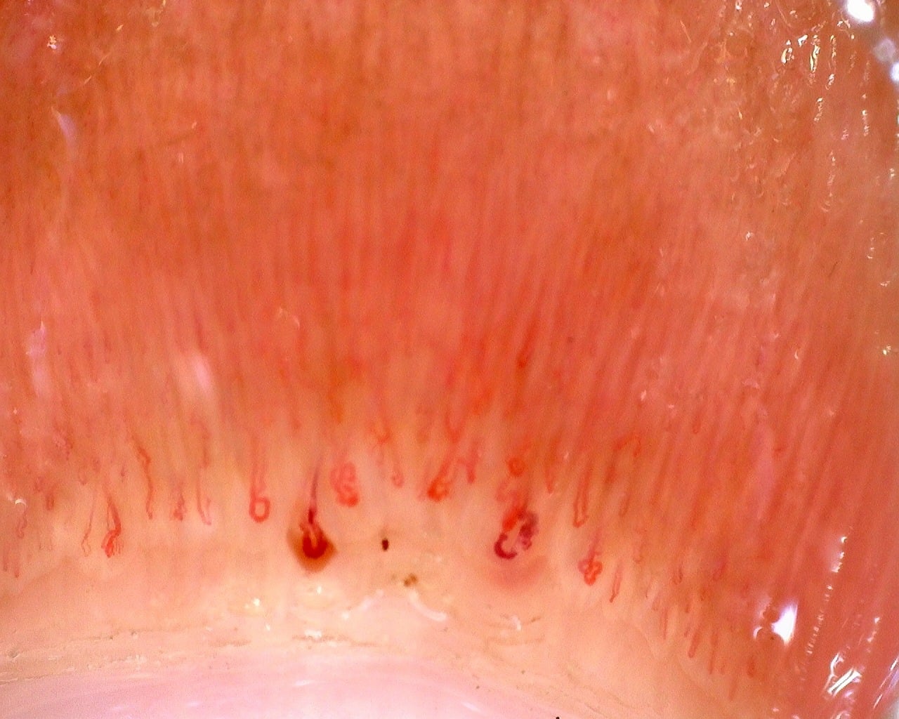
JDM, juvenile dermatomyositis.
Pediatric lupus and other connective tissue diseases
In childhood-onset lupus, findings are less specific. A 2020 systematic review found limited and heterogeneous evidence, with some series reporting hemorrhages, abnormal morphology, or a minority of scleroderma patterns, but no validated unique pattern [12]. This fits the 2023 international cohort, in which the largest qualitative differences from controls occurred in SSc, JDM, and connective tissue diseases, rather than pediatric primary Raynaud's phenomenon (PRP) or JIA [2].
Capillaroscopy has also attracted interest in pediatric localized scleroderma, particularly for longitudinal comparisons with healthy controls. A 2025 systematic review highlighted increased tortuosity, abnormal shapes, or mild vascular changes as the main findings, but concluded that standardization and demonstrated clinical utility remain insufficient [13]. A systematic review of pediatric autoimmune diseases reached a similar conclusion: the technique is promising, but the strength of evidence varies considerably by condition [14].
| Condition | Main contribution of the technique | Current level of confidence |
|---|---|---|
| Pediatric Raynaud's phenomenon | Follow-up stratification alongside ANA and clinical findings [4, 5, 6, 7] | Moderate |
| jSSc | Detection of the scleroderma pattern and vascular follow-up [8, 9] | High within pediatrics |
| JDM | Vascular activity, density, and changes over time [10, 11] | Moderate to high |
| cSLE | Documentation of abnormalities and support for vascular phenotype characterization [12] | Low to moderate |
| Localized scleroderma | Phenotypic hypotheses and comparison with controls [13] | Exploratory |
Practical image acquisition in children
Pediatric capillaroscopy quality depends on both technique and setting. A poor initial experience can make cooperation more difficult throughout the examination [1, 2].
The international multicenter series described above used a protocol of 16 images per patient, covering the second to fifth fingers of both hands [2]. This reflects a practical reality: in children, a somewhat shorter but consistent protocol may be preferable to exhaustive acquisition of poor-quality images. The objective is the same as in adults, but tolerance differs.
- Explain the test before starting and show an image to reduce anxiety.
- Prioritize thermal comfort and sufficient acclimatization; a cold or uncomfortable child cooperates less well and tends to withdraw the hand.
- Avoid excessive pressure from the lens, because compression more readily obscures the distal row in small fingers.
- If the child cannot tolerate a long protocol, obtain at least clear, comparable images of the most evaluable fingers.
- Record nail biting, cuticle trauma, or poor cooperation, because these strongly influence interpretation.
Short cases
Case 1. An adolescent with Raynaud's phenomenon and positive ANA
An adolescent girl has triphasic episodes, ANA at 1:320, and capillaroscopy showing some dilated capillaries without clear capillary loss. The finding does not identify a specific connective tissue disease, but justifies closer follow-up than in ANA-negative pediatric Raynaud's phenomenon with entirely normal capillaroscopy [4, 5, 6, 7].
Case 2. A child with active JDM
A child with proximal weakness, rash, and elevated muscle enzymes has capillary rarefaction and increased diameters. Capillaroscopy supports active vasculopathy and may serve as a complementary biomarker during follow-up, alongside muscle strength, enzyme levels, and skin assessment [10, 11].
FAQ
Is normal capillaroscopy in children the same as in adults?
Not entirely. Some mild morphological variants are more frequent and require greater caution [2, 3].
When is capillaroscopy most useful in pediatrics?
Mainly in Raynaud's phenomenon, suspected jSSc, JDM, and some overlap connective tissue diseases [1, 2, 4, 8, 9, 10, 11].
Does abnormal capillaroscopy in pediatric Raynaud's phenomenon predict progression?
It raises suspicion and may change follow-up, but cannot predict individual progression accurately enough on its own [5, 6, 7].
Which pediatric diseases show the strongest capillaroscopic findings?
jSSc and JDM are currently the settings in which capillaroscopy adds the most [8, 9, 10, 11].
Is it useful in pediatric lupus?
It may detect abnormalities and support vascular characterization, but specificity remains limited [12].
Glossary
- Pediatric Raynaud's phenomenon
- Raynaud's phenomenon in children or adolescents; its prognostic significance depends on clinical findings, serology, and longitudinal follow-up.
- jSSc
- Juvenile systemic sclerosis, one of the pediatric conditions in which capillaroscopy has the greatest diagnostic and follow-up value.
- JDM
- Juvenile dermatomyositis, an inflammatory disease in which capillary loss and diameter changes may accompany vascular activity.
- Capillary rarefaction
- A reduction in the number of visible capillaries per millimeter; in pediatrics, it carries more weight when clear and reproducible.
- PRP
- Pediatric primary Raynaud's phenomenon, in which capillaroscopy is usually normal or nonspecific and is used mainly for follow-up.
References
- Schonenberg-Meinema D, Cutolo M, Smith V. Capillaroscopy in the daily clinic of the pediatric rheumatologist. Best Pract Res Clin Rheumatol. 2024;38(3):101978. DOI: 10.1016/j.berh.2024.101978. PMID: 39034217.
- Melsens K, Cutolo M, Schonenberg-Meinema D, Foeldvari I, Leone MC, Mostmans Y, et al. Standardized nailfold capillaroscopy in children with rheumatic diseases: a worldwide study. Rheumatology (Oxford). 2023;62(4):1605-1615. doi: 10.1093/rheumatology/keac487. PMID: 36005889.
- Terreri MT, Andrade LE, Puccinelli ML, Hilário MO, Goldenberg J. Nail fold capillaroscopy: normal findings in children and adolescents. Semin Arthritis Rheum. 1999;29(1):36-42. doi: 10.1016/S0049-0172(99)80036-5. PMID: 10468413.
- Pain CE, Constantin T, Toplak N, Moll M, Iking-Konert C, Piotto DP, et al; Paediatric Rheumatology European Society (PRES) Juvenile Scleroderma Working Group. Raynaud's syndrome in children: systematic review and development of recommendations for assessment and monitoring. Clin Exp Rheumatol. 2016;34 Suppl 100(5):200-206. PMID: 27494080.
- Piotto DGP, Hilário MOE, Carvalho NS, Len CA, Andrade LEC, Terreri MTRA. Prospective nailfold capillaroscopy evaluation of Raynaud's phenomenon in children and adolescents. Acta Reumatol Port. 2013;38(2):114-121. PMID: 24141348.
- Farenhorst CA, van Roon AM, van Gessel AI, Stel AJ, Bootsma H, Armbrust W, et al. Capillary microscopy is a potential screening method for connective tissue disease in children with Raynaud's phenomenon. Pediatr Rheumatol Online J. 2022;20(1):11. doi: 10.1186/s12969-022-00671-0. PMID: 35135560.
- Mueller M, Gschwandtner ME, Emminger W, Kiener H, Schnaubelt S, Giurgea GA, et al. Associations between nailfold capillary aberrations and autoantibodies in children and adults with Raynaud's phenomenon. RMD Open. 2023;9(1):e003077. doi: 10.1136/rmdopen-2023-003077. PMID: 36972928.
- Foeldvari I, Culpo R, Sperotto F, Anton J, Avcin T, Baildam E, et al. Consensus-based recommendations for the management of juvenile systemic sclerosis. Rheumatology (Oxford). 2021;60(4):1651-1658. doi: 10.1093/rheumatology/keaa584. PMID: 33147624.
- Ingegnoli F, Boracchi P, Gualtierotti R, Smith V, Cutolo M, Foeldvari I; EUSTAR co-authors. A comparison between nailfold capillaroscopy patterns in adulthood in juvenile and adult-onset systemic sclerosis: A EUSTAR exploratory study. Microvasc Res. 2015;102:19-24. doi: 10.1016/j.mvr.2015.07.007. PMID: 26254260.
- Pachman LM, Morgan G, Klein-Gitelman MS, Ahsan N, Khojah A. Nailfold capillary density in 140 untreated children with juvenile dermatomyositis: an indicator of disease activity. Pediatr Rheumatol Online J. 2023;21(1):118. DOI: 10.1186/s12969-023-00903-x. PMID: 37828536.
- Bica PF, Hysa E, Campitiello R, Sulli A, Pizzorni C, Gotelli E, et al. Nailfold videocapillaroscopy in juvenile dermatomyositis: detailed correlations between microvascular abnormalities with clinical and laboratory parameters from an observational retrospective single-center study. Clin Rheumatol. 2025;44(9):3615-3627. doi: 10.1007/s10067-025-07561-2. PMID: 40690123.
- Schonenberg-Meinema D, Melsens K, Nassar-Sheikh Rashid A, Cutolo M, Kuijpers TW, van den Berg JM, et al. Capillaroscopy in childhood-onset systemic lupus erythematosus: a first systematic review. Clin Exp Rheumatol. 2020;38(2):350-354. doi: 10.55563/clinexprheumatol/azl2bj. PMID: 31858964.
- Taşkın SN, Doğantan Ş, Esen E, Özdemir Çiçek S, Paç Kısaarslan A, Poyrazoğlu MH. Revealing Microvascular Involvement in Pediatric Localized Scleroderma Through Nailfold Capillaroscopy. Children (Basel). 2025;12(9):1245. doi: 10.3390/children12091245. PMID: 41007110.
- Li AR, Burke CD, Purvis CS, Lee LW. Association between nailfold capillaroscopy abnormalities and autoimmune disease in pediatric populations. Pediatr Dermatol. 2022;39(2):197-204. doi: 10.1111/pde.14926. PMID: 35078273.
- McClellan N, Chen L, Matossian S, et al. Nailfold capillary phenotypes distinguish juvenile myositis subtypes and associate with disease activity. Clin Transl Med. 2026;16(9):e70789. doi: 10.1002/ctm2.70789. PMID: 42681940.