In inflammatory myopathies, capillaroscopy becomes more informative when interpreted alongside myositis-specific autoantibodies. Anti-MDA5 (melanoma differentiation-associated gene 5), anti-TIF1gamma, anti-Mi2, and some antisynthetase phenotypes show microvascular findings of interest. However, most studies remain small and heterogeneous, so they do not establish the technique as a standalone prognostic biomarker [1, 2, 3, 4, 5, 6, 7, 8, 9, 10, 11].
- Serology and capillaroscopy complement each other. Autoantibodies help define the clinical phenotype, while capillaroscopy provides information on microvascular burden and vascular activity [1, 2, 3, 4].
- The most consistent capillaroscopic findings occur with anti-MDA5, anti-TIF1gamma, and, in some cohorts, anti-Mi2, with more dilations, hemorrhages, or scleroderma-like patterns than in other subgroups [2, 3, 4, 5, 6, 7]. In antisynthetase syndrome, the main contribution is identifying heterogeneity among anti-ARS subtypes rather than suggesting a single pattern [10, 11].
- In anti-MDA5-positive patients, several studies suggest that hemorrhages, disorganization, and certain capillaroscopic scores relate to activity and the pulmonary phenotype, but no prognostic threshold has yet been validated for clinical practice [4, 5, 6, 7].
- The association between anti-TIF1gamma and microangiopathy appears real, but its value for detecting associated cancer remains uncertain; current evidence does not justify using capillaroscopy for cancer screening [2, 3, 8].
- Longitudinal changes appear to differ by autoantibody, supporting the view that myopathies do not all share the same vascular behavior [4, 9, 11].
- The main practical rule is simple: no capillaroscopic pattern should displace standard interstitial lung disease (ILD) or cancer screening, lung function testing, or systemic activity assessment [1, 4, 5, 6, 7, 8, 9, 10].
Learning path
If you already know the serological profile and want to understand what capillaroscopy adds, this unit covers its practical value in anti-MDA5, anti-TIF1gamma, and antisynthetase syndromes. The aim is to distinguish interesting associations that do not change management from information that supports specific clinical actions [1, 2, 3, 4, 5, 6, 7, 8, 9, 10, 11].
What capillaroscopy adds to serology
Myositis-specific autoantibodies already shape much of the clinical phenotype. The practical question is whether capillaroscopy adds useful information on vascular burden, activity, or the distribution of damage, rather than whether it replaces serology [1, 2, 3, 4].
Pauling et al's review of vasculopathy in inflammatory myopathies and Flatley et al's systematic review agree that microangiopathy is not an irrelevant secondary finding, particularly in dermatomyositis. Interpretation nevertheless requires standardized language and recognition that most cohorts are small [1, 2]. The multicenter cohort by Torres-Ruiz et al reinforces this view: the scleroderma pattern occurred mainly in dermatomyositis and overlap syndromes, whereas nonspecific patterns were more common in antisynthetase syndrome or phenotypes without dermatomyositis [3].
In practice, capillaroscopy may help answer four questions: whether vasculopathy is severe or mild, whether the phenotype appears more inflammatory or more fibrosing, whether microangiopathy improves with treatment, and whether the capillary pattern is consistent with or discordant with the specific serological profile [1, 2, 3, 4, 9, 10]. It still cannot reliably predict on its own who will develop rapidly progressive ILD, who will have cancer, or who will respond best to a particular therapy.
| Autoantibody / group | Reported capillary findings | Current utility | Limitation |
|---|---|---|---|
| Anti-MDA5 | More hemorrhages, disorganization, and dynamic changes [4, 5, 6, 7] | Helps contextualize activity and the pulmonary phenotype | No validated prognostic cutoffs |
| Anti-TIF1gamma | More abnormal patterns in some cohorts [3, 4, 8] | Suggests a high microvascular burden | Cannot provide cancer screening on its own |
| Anti-Mi2 | Scleroderma-pattern features or avascular areas in some series [3, 4] | Supports phenotype characterization | Small samples |
| Antisynthetase / anti-Jo1 | More heterogeneous, with dilations, hemorrhages, or fewer visible capillaries in some cases [3, 10, 11] | Complementary stratification | Lower specificity than in dermatomyositis |
Anti-MDA5 and the pulmonary phenotype
The anti-MDA5 subgroup has driven much of the recent capillaroscopy research because it combines a striking vascular phenotype with a high clinical risk of rapidly progressive ILD [4, 5, 6, 7].
In the cohort by Kubo et al, 87.5% of anti-MDA5-positive patients had capillaroscopic abnormalities [4]. Flatley et al also found a clear signal in their dermatomyositis review: 15 of 31 patients with anti-MDA5 had a scleroderma-like pattern, and 13 had hemorrhages and dilation [2]. These figures are not definitive, but are consistent enough to suggest that anti-MDA5 serology increases the relevance of capillaroscopy, and vice versa.
The study by Sugimoto et al is particularly relevant because it prospectively followed anti-MDA5-positive patients with dermatomyositis and reported three useful findings: improvement in the capillary score with immunosuppression, a positive correlation with forced vital capacity, and an inverse correlation with anti-MDA5 titers [5]. The cautious interpretation is that visible microangiopathy does not always mean more severe lung damage; in some cases, it may reflect a more cutaneous–vascular phenotype rather than fulminant pulmonary involvement. This hypothesis still requires confirmation.
Wakura compared anti-MDA5 and antisynthetase antibodies in patients with dermatomyositis complicated by ILD and found more hemorrhages and disorganization with anti-MDA5. Capillary loss was associated with pulmonary fibrosis, while hemorrhages were associated with more aggressive activity [6]. Tang extended this work in 2025 by associating certain capillaroscopic abnormalities with interferon activation and cytokine profiles, adding biological plausibility, although the findings remain exploratory [7].
Anti-TIF1gamma, anti-Mi2, and cancer risk
Anti-TIF1gamma and, to a lesser extent, anti-Mi2 are the other antibody groups that have received most attention for their possible association with more pronounced capillaroscopic findings [2, 3, 4, 8].
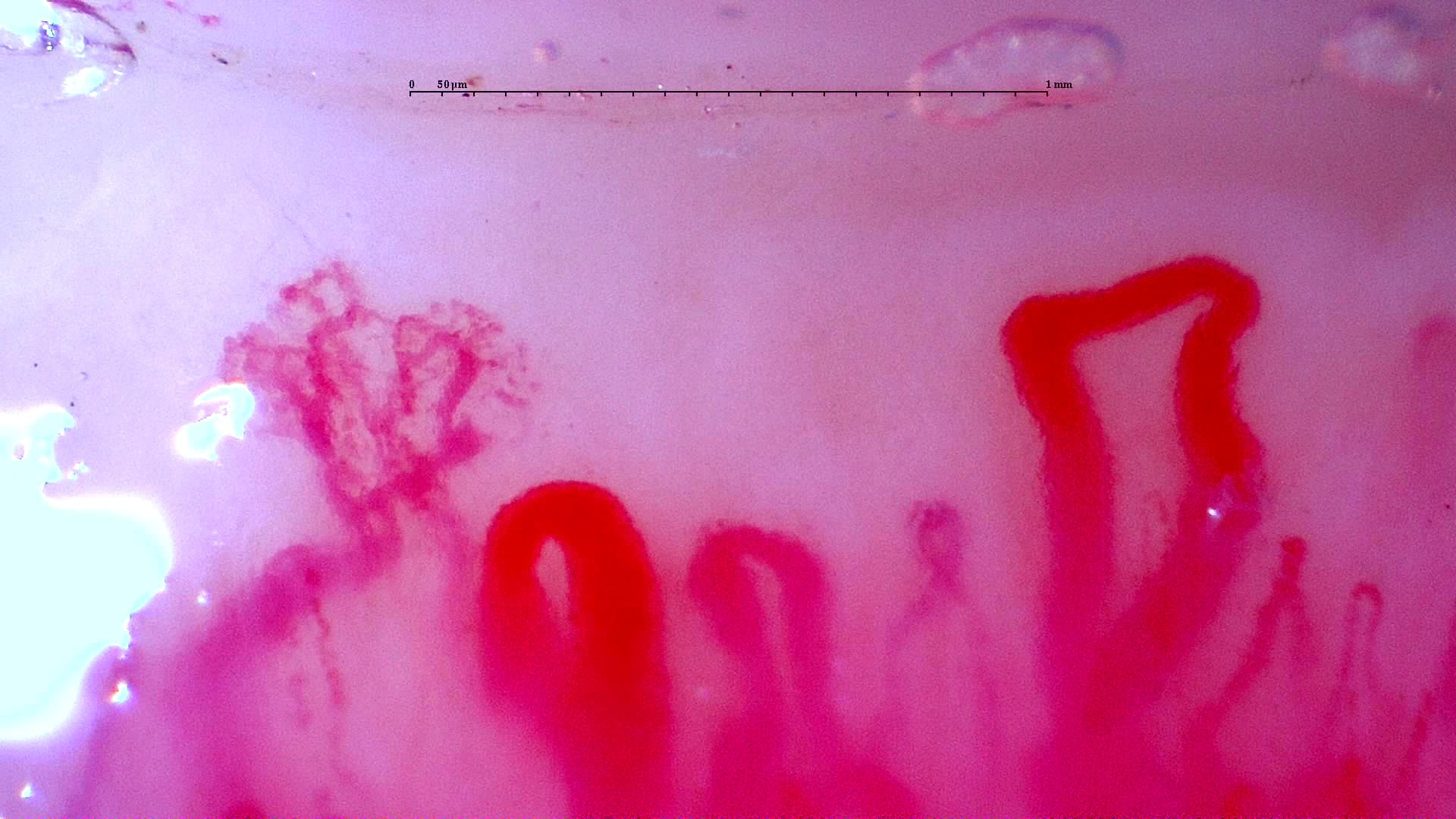
In Flatley et al's systematic review, 8 of 10 patients with anti-TIF1gamma had a scleroderma-like pattern, a clear signal of overt microangiopathy [2]. Torres-Ruiz et al also identified an association between the scleroderma pattern and anti-TIF1gamma in a multiethnic cohort [3]. Anti-Mi2 data are more modest, but point in the same general direction: a greater burden of capillary abnormalities than in other myopathy subgroups [2, 3, 4].
The difficulty arises when these findings are treated as a cancer marker. Mumtaz et al found greater density loss and more hemorrhages in anti-TIF1gamma dermatomyositis than in other myopathy subtypes, but no statistically significant differences between anti-TIF1gamma-positive patients with and without cancer [8]. Capillaroscopy may therefore suggest marked microangiopathy in a serological context associated with risk, but it does not remove the need for cancer screening.
Insufficient evidence / not found. There is insufficient evidence to use capillaroscopy to screen for occult cancer in anti-TIF1gamma dermatomyositis.
Antisynthetase syndrome and other subgroups
Compared with anti-MDA5- or anti-TIF1gamma-associated disease, antisynthetase syndrome usually has less striking and more heterogeneous capillaroscopic findings [3, 4, 6, 10, 11].
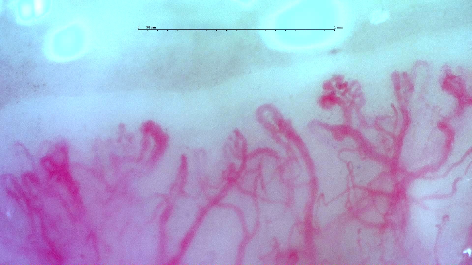
In Sebastiani's study, a scleroderma-like pattern occurred in 35.3% of antisynthetase syndrome cases, while any abnormality was present in 62.1% [10]. Kubo et al had already observed fewer abnormalities in the anti-ARS group than in the anti-MDA5 or anti-TIF1gamma groups [4]. In practice, capillaroscopy may support an antisynthetase phenotype, but will rarely be the decisive diagnostic test.
A recent study by Mugii et al added a clinically useful qualification: anti-ARS subtypes do not all behave alike within the antisynthetase spectrum. Among 66 Japanese patients, dilations, fewer visible capillaries, and hemorrhages were frequent, whereas complex abnormal shapes, disorganization, and capillary loss were rare. The anti-KS subtype also had milder microvascular involvement than the others [11]. The article's abstract uses the classical term capillary ramifications [11]; here, we use the preferred category of abnormal shapes.
Longitudinal reversibility also appears to depend on the autoantibody. Mugii et al had previously described different trajectories across serological groups in a longitudinal cohort, reinforcing that microangiopathy in myositis does not follow a single course [9]. Follow-up should therefore be interpreted in the context of serology as well as the overall clinical diagnosis.
Using capillaroscopy without overstating the evidence
The clinical challenge is to distinguish association, which may be interesting and informative without changing management, from evidence that supports specific actions. Serological associations are of interest; evidence for concrete management strategies remains more limited [1, 2, 3, 4, 5, 6, 7, 8, 9, 10, 11].
- Use capillaroscopy to contextualize serology, not to replace it.
- Describe specific domains before assigning prognostic meaning.
- In anti-MDA5-positive patients, relate the pattern to vascular activity without presenting a definitive pulmonary prognosis.
- In anti-TIF1gamma-positive patients, avoid wording that suggests cancer screening should be guided by capillaroscopy.
- If the capillaroscopic pattern changes during follow-up, compare it with treatment, lung function, ferritin, creatine kinase (CK), and extramuscular activity.
Short cases
Case 1. Anti-MDA5 dermatomyositis with respiratory deterioration
An anti-MDA5-positive woman has abundant hemorrhages and capillary disorganization. The finding raises suspicion of substantial vascular activity, but the critical decision remains urgent completion of pulmonary assessment and adjustment of treatment to the actual respiratory phenotype [4, 5, 6, 7].
Case 2. Anti-TIF1gamma dermatomyositis and cancer investigation
Capillaroscopy shows rarefaction and hemorrhages. The pattern supports an active microvascular phenotype, but does not change the central point: cancer screening should follow the serological profile and clinical assessment, rather than the capillary image [2, 3, 8].
FAQ
Which autoantibody has been studied most in relation to capillaroscopy?
Anti-MDA5, followed by anti-TIF1gamma; data are also available for anti-Mi2 [2, 3, 4, 5, 6, 7, 8].
Does a high capillary score in anti-MDA5 disease mean more severe lung damage?
Not necessarily. Some data suggest an inverse relationship with antibody titers and an association with better lung function. In any case, the evidence remains insufficient to use this as an individual prognostic rule [5].
Can capillaroscopy replace ferritin measurement or lung function testing in anti-MDA5 disease?
No. It should be considered complementary information [4, 5, 6, 7].
Can it screen for cancer in anti-TIF1gamma disease?
Do findings change over time according to the autoantibody?
Probably yes. Available longitudinal data suggest different trajectories by serological group, as well as heterogeneity among anti-ARS subtypes [4, 9, 11].
What should the report include?
A structured description of domains, the known serological phenotype, and a cautious conclusion about microvascular activity or the predominant pattern, without claiming definitive prognostic ability.
Glossary
- MSA
- Myositis-specific autoantibodies, useful for identifying clinical phenotypes and predicting the risk of extramuscular involvement.
- Anti-MDA5
- Autoantibodies associated with amyopathic or hypomyopathic dermatomyositis and a high risk of rapidly progressive ILD.
- Anti-TIF1gamma
- Dermatomyositis autoantibodies associated with increased cancer risk, particularly in adults.
- Hemorrhages
- Extravasations visible on capillaroscopy that may indicate recent or active microvascular damage.
- Rapidly progressive pulmonary phenotype
- An acute or subacute ILD course with rapidly worsening symptoms, lung function, and chest imaging.
References
- Pauling JD, Christopher-Stine L. The aetiopathogenic significance, clinical relevance and therapeutic implications of vasculopathy in idiopathic inflammatory myopathy. Rheumatology (Oxford). 2021;60(4):1593-1607. doi: 10.1093/rheumatology/keaa816. PMID: 33458769.
- Flatley EM, Collins D, Lukowiak TM, Miller JH. Nailfold microscopy in adult-onset dermatomyositis in association with myositis antibodies. Arch Dermatol Res. 2024;317(1):34. doi: 10.1007/s00403-024-03521-z. PMID: 39565371.
- Torres-Ruiz J, Pinal-Fernandez I, Selva-O'Callaghan A, Campbell B, Muñoz-Braceras S, Mejía-Domínguez NR, et al. Nailfold capillaroscopy findings of a multicentric multi-ethnic cohort of patients with idiopathic inflammatory myopathies. Clin Exp Rheumatol. 2024;42(2):367-376. doi: 10.55563/clinexprheumatol/l9gudh. PMID: 38488092.
- Kubo S, Todoroki Y, Nakayamada S, Nakano K, Satoh M, Nawata A, et al. Significance of nailfold videocapillaroscopy in patients with idiopathic inflammatory myopathies. Rheumatology (Oxford). 2019;58(1):120-130. doi: 10.1093/rheumatology/key257. PMID: 30169697.
- Sugimoto T, Mokuda S, Kohno H, Ishitoku M, Araki K, Watanabe H, et al. Nailfold capillaries and myositis-specific antibodies in anti-melanoma differentiation-associated gene 5 antibody-positive dermatomyositis. Rheumatology (Oxford). 2022;61(5):2006-2015. doi: 10.1093/rheumatology/keab681. PMID: 34498053.
- Wakura R, Matsuda S, Kotani T, Shoda T, Takeuchi T. The comparison of nailfold videocapillaroscopy findings between anti-melanoma differentiation-associated gene 5 antibody and anti-aminoacyl tRNA synthetase antibody in patients with dermatomyositis complicated by interstitial lung disease. Sci Rep. 2020;10(1):15692. doi: 10.1038/s41598-020-72752-7. PMID: 32973255.
- Tang M, Shi J, Pang Y, Zhou S, Liu J, Wu C, et al. Nailfold videocapillaroscopy findings are associated with IIM subtypes and IFN activation. Arthritis Res Ther. 2025;27(1):62. doi: 10.1186/s13075-025-03532-9. PMID: 40114239.
- Mumtaz S, Phillipps J, Sullivan MM, Diaz-Menindez M, Wang B, Majithia V, et al. Microvascular abnormalities between anti-TIF1-γ-associated dermatomyositis with and without malignancy. BMC Rheumatol. 2025;9(1):50. doi: 10.1186/s41927-025-00504-z. PMID: 40329336.
- Mugii N, Hamaguchi Y, Horii M, Fushida N, Ikeda T, Oishi K, et al. Longitudinal changes in nailfold videocapillaroscopy findings differ by myositis-specific autoantibody in idiopathic inflammatory myopathy. Rheumatology (Oxford). 2023;62(3):1326-1334. doi: 10.1093/rheumatology/keac401. PMID: 35866689.
- Sebastiani M, Triantafyllias K, Manfredi A, González-Gay MA, Palmou-Fontana N, Cassone G, et al. Nailfold Capillaroscopy Characteristics of Antisynthetase Syndrome and Possible Clinical Associations: Results of a Multicenter International Study. J Rheumatol. 2019 Mar;46(3):279-284. doi: 10.3899/jrheum.180355. Epub 2018 Nov 15. PMID: 30442823.
- Mugii N, Hamaguchi Y, Fushida N, Fujii K, Nishio J, Kudo K, et al. Distinct Nailfold Videocapillaroscopy Findings in Patients With Anti-Aminoacyl-tRNA Synthetase Antibodies. J Dermatol. 2026;53(2):318-322. doi: 10.1111/1346-8138.70081. PMID: 41312673.