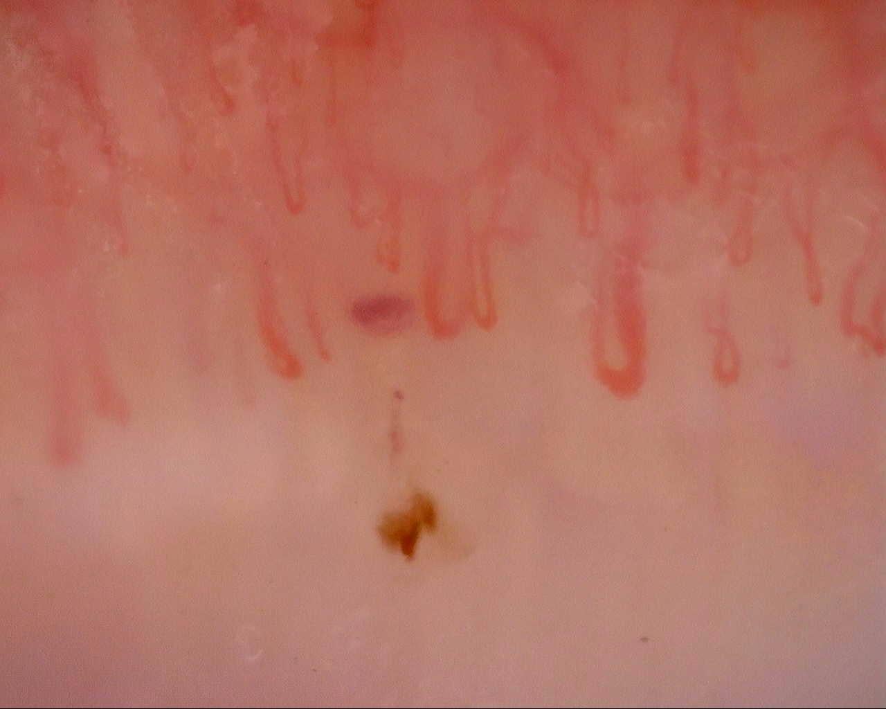
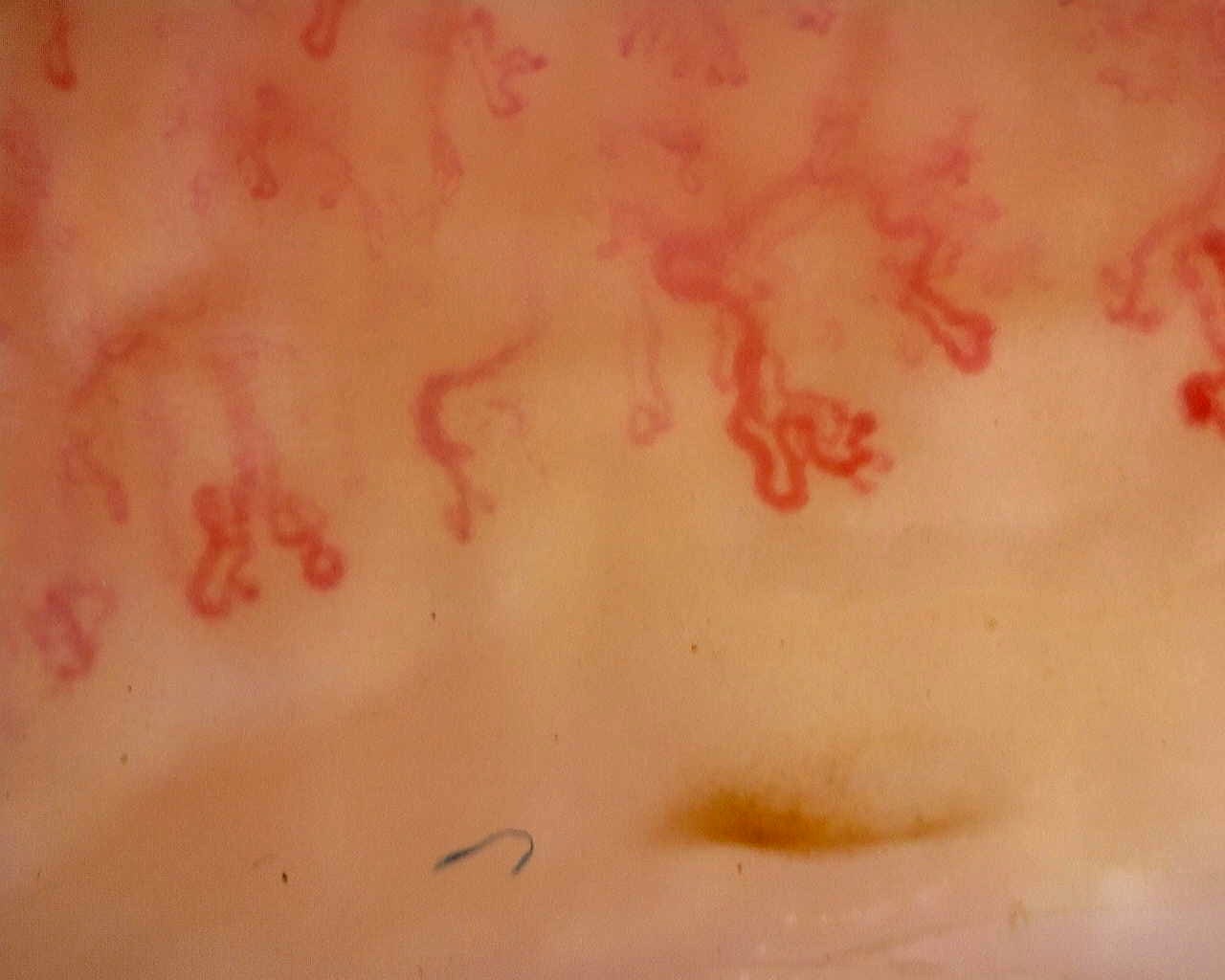
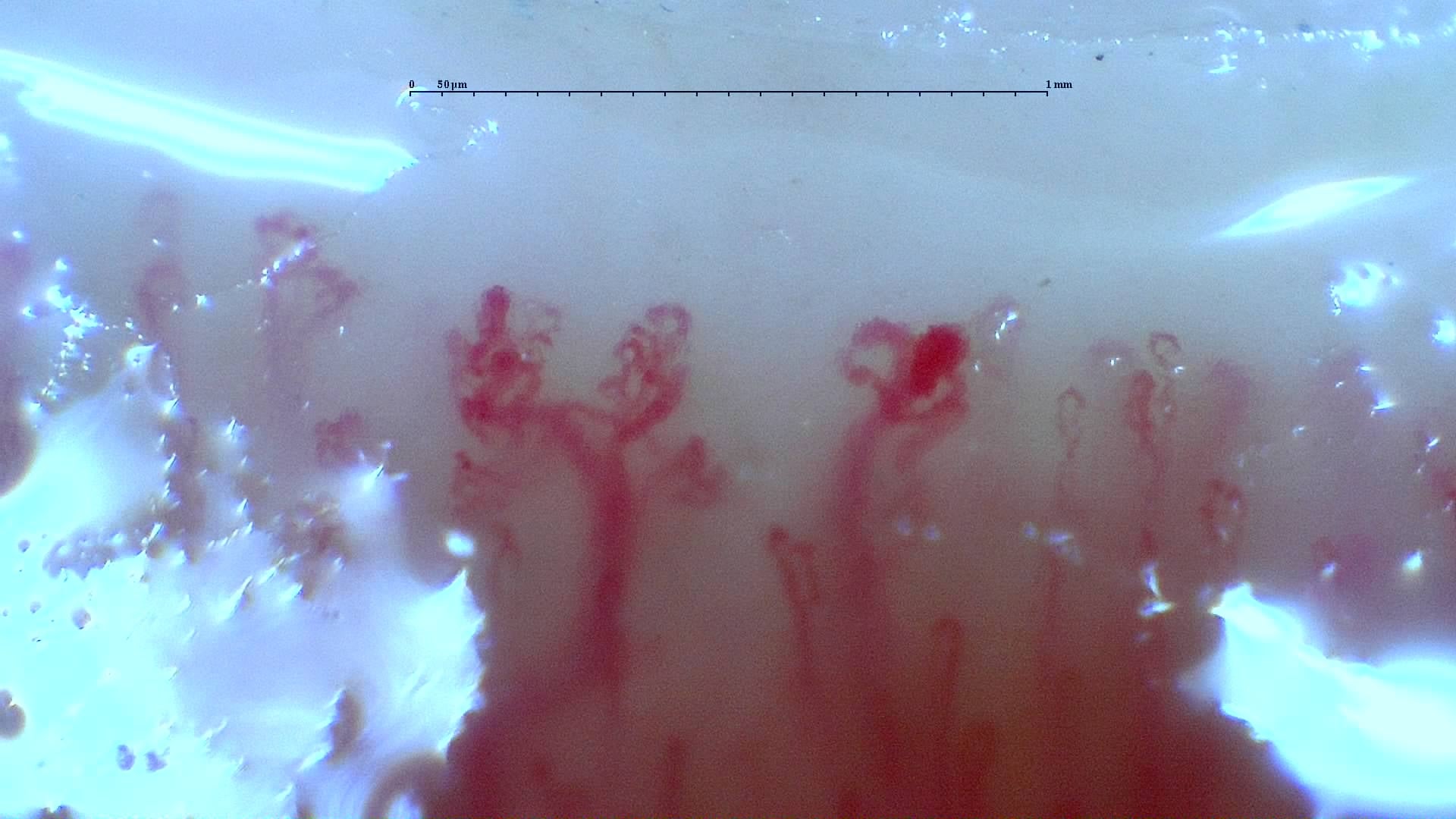
In MCTD, capillaroscopy helps identify microangiopathy within the scleroderma spectrum, but interpretation requires greater caution than in systemic sclerosis. The scleroderma-like pattern is frequent, may change over time, and has been associated with pulmonary hypertension, sclerodactyly, and interstitial involvement in several cohorts, although the evidence remains observational and heterogeneous [1, 2, 3, 4, 5, 6, 7, 8, 9].
- MCTD is an overlap disease characterized by anti-U1-RNP antibodies and features of lupus, systemic sclerosis, and myositis; capillaroscopy must therefore always be interpreted within a broad clinical context [1, 2, 3].
- Recent cohorts show a clear scleroderma pattern in 38% to 46.5% of patients. When nonspecific microangiopathy is included, abnormal examinations readily account for more than half of patients [4, 5, 6, 7].
- The early phase is the most frequent; an advanced or highly destructive appearance should prompt reassessment of whether the phenotype is closer to systemic sclerosis than to “pure” MCTD [4, 5, 6, 7].
- In prospective and retrospective studies, severe capillary disorganization has been associated with interstitial lung involvement, and the scleroderma pattern with pulmonary arterial hypertension [6, 7, 8].
- Capillaroscopic findings appear more dynamic in MCTD than in systemic sclerosis: some patients show normalization and others improve with immunosuppression, which is unusual in the classical scleroderma spectrum [7, 9].
- A scleroderma-like pattern in MCTD does not reclassify the case as systemic sclerosis, but it does indicate a vascular phenotype with greater prognostic relevance that warrants closer follow-up [1, 2, 6, 7, 8].
Learning path
This unit provides the key context for interpreting a scleroderma-like pattern without automatically assuming systemic sclerosis. You may then find it useful to review dermatomyositis and inflammatory myopathies, where the same pattern appears with a different clinical interpretation [1, 2, 4, 5, 6, 7, 8, 9].
Why capillaroscopy matters in MCTD
MCTD is defined by anti-U1-RNP autoantibodies and a variable combination of manifestations resembling lupus, systemic sclerosis, or myositis. This mixed nature explains both the value of capillaroscopy and why its interpretation should be less rigid than in the classical systemic sclerosis algorithm [1, 2, 3].
In practice, capillaroscopy serves three purposes. First, it supports differential diagnosis when a patient has Raynaud's phenomenon, swollen hands, synovitis, or myositis and does not yet clearly fit a specific connective tissue disease [1, 2, 3]. Second, it identifies a microvascular phenotype closer to the scleroderma spectrum, particularly when giant capillaries, avascular areas, or capillary rarefaction are present [4, 5, 6, 7, 8]. Third, it helps monitor vascular and pulmonary complications, although the evidence here remains moderate and mainly observational [6, 7, 8, 9].
The technique is also useful because it requires two distinct issues to be considered separately. A capillary finding may strongly suggest structural microvascular damage without necessarily determining the final disease classification. This matters in MCTD, where disease can evolve over time and some cases are reclassified as lupus, systemic sclerosis, or more clearly defined overlap syndromes [1, 2, 3].
Patterns and frequency
Abnormal capillaroscopy is frequent in MCTD, but the exact proportion depends on how abnormalities are defined, the number of fingers examined, treatment received, and whether a complete scleroderma pattern is required or nonspecific microangiopathy is also included [4, 5, 6, 7, 8, 9].
Recent cohorts report a scleroderma pattern in 38% to 46.5% of cases when strict definitions are used [4, 7]. Including nonspecific findings or early scleroderma-like changes clearly increases the proportion of abnormal patterns. In the cohort reported by Felis-Giemza et al, for example, up to 69.6% of 79 patients had capillaroscopic changes, with the early pattern, present in 51.9% of cases, being the most frequent [5]. In the series by Kasser et al, three patterns coexisted among 40 evaluable patients: normal in 15%, nonspecific microangiopathy in 40%, and a scleroderma pattern in 45% [6].
The predominance of the early pattern makes clinical sense. MCTD may share giant capillaries, dilations, and hemorrhages with systemic sclerosis, but does not always develop the same destructive rarefaction or the same longitudinal pattern stability [4, 5, 6, 7, 8, 9]. When capillary loss is very marked or the appearance clearly suggests a late pattern, reassess whether the case is evolving toward a more scleroderma-dominant phenotype [1, 2, 6].
| Study | Population | Main findings | Practical interpretation |
|---|---|---|---|
| Ornowska et al, 2024 [4] | 43 patients with MCTD | Scleroderma-like pattern in 46.5%, predominantly the early phase | The scleroderma pattern is frequent, but not universal |
| Felis-Giemza et al, 2022 [5] | 79 patients with MCTD | Changes in 69.6%; early phase in 51.9% | Abnormalities are more common than fully developed patterns |
| Kasser et al, 2025 [6] | 40 evaluable examinations | 15% normal, 40% nonspecific, 45% scleroderma pattern | Distinguish nonspecific microangiopathy from a true scleroderma pattern |
| Todoroki et al, 2022 [7] | 50 newly diagnosed, untreated cases | Scleroderma pattern in 38% | Prevalence falls when a stricter pattern definition is required |
| de Holanda et al, 2007 [9] | 63 patients with follow-up | Scleroderma (SD) pattern in 65% at baseline, with longitudinal changes in 16% | Findings may improve or normalize in a meaningful minority |
Associations with organ involvement
The main current interest in capillaroscopy in MCTD is its contribution to stratifying phenotypes with a greater vascular or pulmonary burden, rather than independently “confirming” the disease [4, 5, 6, 7, 8, 9].
Pulmonary arterial hypertension
The best-described association is with pulmonary arterial hypertension. In the Japanese prospective multicenter cohort of 50 untreated patients, 22% had pulmonary arterial hypertension, and all of these patients had a scleroderma pattern; among those without pulmonary hypertension, pattern prevalence was 21% [7]. The series by Ornowska et al also found an association between abnormal shapes, a visible subpapillary plexus, and pulmonary hypertension [4]. These findings are consistent, but come from small cohorts. Capillaroscopy may therefore raise suspicion, but it does not replace echocardiography or hemodynamic assessment when indicated [4, 7].
Interstitial lung disease
Interstitial involvement also appears to be related to capillary loss. In the study by Kasser et al, patients with interstitial lung disease (ILD) had fewer capillaries per millimeter, and those with density ≤4/mm had a higher likelihood of ILD, even independently of skin sclerosis or an overall scleroderma pattern [6]. The series by Celińska-Löwenhoff et al also identified giant capillaries as an independent factor associated with pulmonary fibrosis in a cohort of 30 patients with very early disease [8]. These data are clinically useful, but do not yet justify using capillaroscopy as a standalone lung screening test [6, 8].
Skin and the scleroderma phenotype
Sclerodactyly and skin sclerosis are among the features most commonly associated with the scleroderma pattern and capillary rarefaction [4, 5, 6]. Capillary findings are not simply a reflection of the skin: in some cohorts, the relationship with the type of skin lesion was not direct, so a patient with lupus-like skin features could still have scleroderma-type microangiopathy [5].
| Capillary finding | Reported association | Strength of evidence |
|---|---|---|
| Overall scleroderma pattern | Higher prevalence of pulmonary hypertension [4, 7] | Moderate, small cohorts |
| Density ≤4/mm and severe capillary loss | More ILD and greater risk of a pulmonary phenotype [6] | Moderate |
| Giant capillaries | Association with pulmonary fibrosis in early disease [8] | Low to moderate |
| Abnormal vessels / visible plexus | Signal for pulmonary hypertension in one cohort [4] | Exploratory |
Differential diagnosis and overlap syndromes
The main conceptual error in this setting is treating the scleroderma pattern as exclusive to systemic sclerosis. Similar morphology can occur in overlap syndromes, MCTD, and some inflammatory myopathies, but with different frequencies, dynamics, and serological profiles [1, 2, 3, 4, 5, 6, 7].
Capillaroscopy is diagnostically useful when the clinical picture is still incomplete. In a patient with Raynaud's phenomenon, hand edema, arthritis or myositis, and positive anti-U1-RNP antibodies, but no clearly scleroderma-dominant phenotype, giant capillaries, hemorrhages, and capillary loss provide additional diagnostic coherence [1, 2, 3, 4]. These observations do not replace classification criteria, but favor an overlap syndrome with a structural vascular component.
The test also helps avoid overdiagnosis. Nonspecific microangiopathy without clear rarefaction, repeated giant capillaries, or a compatible serological profile should carry less weight than a true scleroderma-like pattern reproducible across several fingers. This distinction matters in MCTD because some patients retain mixed features for years without behaving clinically like overt systemic sclerosis [1, 2, 6].
| Scenario | What capillaroscopy adds | What capillaroscopy does not determine |
|---|---|---|
| Raynaud's phenomenon + anti-U1-RNP + incomplete clinical picture | Supports an overlap phenotype with microvascular damage [1, 2, 3, 4] | Does not establish the final diagnosis on its own |
| MCTD versus systemic sclerosis | Clarifies the severity and phase of vascular damage [4, 5, 6, 7] | Does not resolve uncertain classification alone |
| MCTD versus lupus or UCTD | A scleroderma pattern increases suspicion of overlap [1, 2, 3] | Serology, muscle, skin, and lung findings must still be considered |
UCTD, undifferentiated connective tissue disease.
Follow-up and limitations
The literature on follow-up in MCTD is much smaller than in systemic sclerosis, but suggests an interesting feature: capillaroscopy may change with treatment and inflammatory activity [7, 9].
The classical study by de Holanda et al showed that ten of 63 patients changed pattern over time and that capillaroscopy “normalized” in seven cases; moderate or severe avascular areas were also less frequent at the final assessment than at the previous one [9]. Todoroki et al found something similar in recent-onset disease: after intensive immunosuppression, the scleroderma pattern disappeared in approximately half of patients with MCTD, whereas it remained unchanged in the systemic sclerosis comparator group [7].
This greater capacity for change is appealing, but does not make the technique a validated activity marker. Large longitudinal studies, standardized repeat-examination protocols, and robust thresholds for improvement or progression in MCTD are still lacking. Repeat testing should therefore address a specific question: whether the vascular phenotype is worsening, whether ILD or pulmonary hypertension has appeared, or whether a treatment change has had a measurable effect in a complex case [6, 7, 8, 9].
Insufficient evidence / not found. There is insufficient support for using capillaroscopy as an isolated marker to guide intensification or withdrawal of immunosuppression in MCTD.
Short cases
Case 1. Raynaud's phenomenon, mild myositis, and anti-U1-RNP
A 34-year-old woman presents with recent-onset Raynaud's phenomenon, hand edema, arthralgia, and mildly elevated creatine kinase (CK). Capillaroscopy shows isolated giant capillaries, punctate hemorrhages, and mild rarefaction. This is insufficient to diagnose MCTD, but supports that the case is more than uncomplicated primary Raynaud's phenomenon and requires follow-up as an overlap connective tissue disease [1, 2, 3, 4].
Case 2. Established MCTD and new dyspnea
In a patient with an established diagnosis, new marked rarefaction, density ≤4/mm, and neoangiogenesis should raise concern about a higher risk of interstitial or pulmonary vascular involvement. The useful next step is to complete pulmonary and hemodynamic assessment, rather than simply repeat capillaroscopy a month later [6, 7, 8].
FAQ
Does MCTD have its own capillaroscopic pattern?
There is no exclusive pattern. The most common findings are a scleroderma-like pattern or nonspecific microangiopathy, predominantly in early phases [4, 5, 6, 7].
Is a scleroderma pattern in MCTD associated with a worse prognosis?
It may suggest a greater vascular burden, especially with capillary loss or features of pulmonary hypertension and lung involvement, but it is insufficient to determine prognosis on its own [6, 7, 8].
Should every patient with MCTD undergo capillaroscopy?
At least one baseline assessment is reasonable in the presence of Raynaud's phenomenon, sclerodactyly, suspected vascular overlap, or a need to stratify pulmonary involvement. Repeat testing depends on the clinical question [1, 6, 7, 8].
Can capillaroscopic findings in MCTD improve with treatment?
Yes. Several small cohorts showed improvement or partial normalization, suggesting greater reversibility than in systemic sclerosis [7, 9].
Glossary
- MCTD
- Mixed connective tissue disease, an overlap connective tissue disease associated with anti-U1-RNP antibodies.
- Scleroderma-like pattern
- A set of capillaroscopic findings resembling the scleroderma pattern, although they do not always clearly fit the classical phases of systemic sclerosis.
- Capillary rarefaction
- A reduction in the number of capillaries per millimeter, useful as a marker of structural microvascular damage.
- Neoangiogenesis
- Abnormal or highly irregular capillaries interpreted as a disorganized attempt at vascular repair.
- Anti-U1-RNP
- Autoantibodies characteristic of MCTD and a central component of its clinical diagnosis.
References
- Chaigne B, Scirè CA, Talarico R, Alexander T, Amoura Z, Avcin T, et al. Mixed connective tissue disease: state of the art on clinical practice guidelines. RMD Open. 2018;4(Suppl 1):e000783. doi: 10.1136/rmdopen-2018-000783. PMID: 30402271.
- Gunnarsson R, Hetlevik SO, Lilleby V, Molberg Ø. Mixed connective tissue disease. Best Pract Res Clin Rheumatol. 2016;30(1):95-111. doi: 10.1016/j.berh.2016.03.002. PMID: 27421219.
- Escolà-Vergé L, Pinal-Fernandez I, Fernandez-Codina A, Callejas-Moraga EL, Espinosa J, Marin A, et al. Mixed Connective Tissue Disease and Epitope Spreading: An Historical Cohort Study. J Clin Rheumatol. 2017;23(3):155-159. doi: 10.1097/RHU.0000000000000500. PMID: 28248800.
- Ornowska S, Wudarski M, Dziewięcka E, Olesińska M. Nailfold capillaroscopy in mixed connective tissue disease patients. Clin Rheumatol. 2024;43(5):1703-1709. doi: 10.1007/s10067-024-06879-7. PMID: 38509242.
- Felis-Giemza A, Ornowska S, Haładyj E, Czuszyńska Z, Olesińska M. Relationship between type of skin lesions and nailfold capillaroscopy pattern in mixed connective tissue disease. Clin Rheumatol. 2022;41(1):281-288. doi: 10.1007/s10067-021-05717-4. PMID: 34370129.
- Kasser C, Boleto G, Allanore Y, Avouac J. Nailfold videocapillaroscopy findings and associations with organ involvement in mixed connective tissue disease. Clin Exp Rheumatol. 2025;43(3):418-424. doi: 10.55563/clinexprheumatol/xshsd7. PMID: 39711361.
- Todoroki Y, Kubo S, Nakano K, Miyazaki Y, Ueno M, Satoh-Kanda Y, et al. Nailfold microvascular abnormalities are associated with a higher prevalence of pulmonary arterial hypertension in patients with MCTD. Rheumatology (Oxford). 2022;61(12):4875-4884. doi: 10.1093/rheumatology/keac165. PMID: 35285493.
- Celińska-Löwenhoff M, Pastuszczak M, Pełka K, Stec-Polak M, Wojas-Pelc A, Musiał J. Associations between nailfold capillaroscopy findings and interstitial lung disease in patients with mixed connective tissue disease. Arch Med Sci. 2019;16(2):297-301. doi: 10.5114/aoms.2018.81129. PMID: 32190139.
- de Holanda Mafaldo Diógenes A, Bonfá E, Fuller R, Correia Caleiro MT. Capillaroscopy is a dynamic process in mixed connective tissue disease. Lupus. 2007;16(4):254-258. doi: 10.1177/0961203307076517. PMID: 17439931.