Diabetes mellitus (DM), particularly type 2 diabetes (T2DM), is associated with a systemic microvascular burden that may also be visible on capillaroscopy. Recent literature has strengthened the association between diabetes and findings such as tortuosity, ectasia, hemorrhages, avascular areas, and neoangiogenesis, and has begun exploring links with retinopathy and nephropathy, supported by machine-learning prediction models. The technique nevertheless remains more promising as a research biomarker and an aid to phenotype characterization than as a standard clinical screening test [1, 2, 3, 4, 5, 6, 7, 8, 9, 10].
- Findings are more consistent in T2DM than in type 1 diabetes (T1DM): a recent meta-analysis found significant associations between diabetes and tortuosity, abnormal morphology, ectasia, bushy capillaries, hemorrhages, and avascular areas, particularly in patients with T2DM [1].
- Another 2025 meta-analysis found that diabetic retinopathy was associated with more giant capillaries, neoangiogenesis, hemorrhages, tortuosity, and avascular areas than diabetes without retinopathy [2].
- Evidence for nephropathy is also growing: albuminuria, reduced estimated glomerular filtration rate (eGFR), and higher-risk Kidney Disease: Improving Global Outcomes (KDIGO) categories have been associated with more tortuosity, abnormal shapes, and abnormal capillaroscopic patterns [3, 4].
- The clinical message should remain cautious: capillaroscopy does not replace fundus examination, albuminuria measurement, eGFR, or standard vascular assessment in people with diabetes [1, 2, 3, 4].
- Outside rheumatology, one of the most promising applications is in research and data science: quantifying large capillaroscopy datasets to explore associations between microvascular phenotypes and diabetic complications [5].
- Insufficient evidence / not found. There is currently insufficient support to recommend capillaroscopy for routine microvascular complication screening in everyone with diabetes.
Learning path
This unit explains what capillaroscopy can currently add as a marker of microangiopathy in diabetes and what remains exploratory. It is especially useful after reviewing AI, quantification, and reproducibility, because quantitative and longitudinal assessment matter more in diabetes than identifying a fixed pattern [1, 2, 3, 4, 5].
Why capillaroscopy is of interest in diabetes
The pathophysiological rationale is strong. Diabetes damages the endothelium, alters perfusion, and promotes microvascular remodeling, making it reasonable to investigate whether some of that damage can be detected at the nailfold [1, 2, 3, 4].
The distinction from systemic sclerosis is essential. In diabetes, the aim is to identify a set of signs of chronic microvascular damage rather than a unique capillaroscopic pattern. This changes how the test is interpreted: the question is whether the microvascular profile is compatible with a greater burden of microangiopathy, rather than whether a “diabetic pattern” is present [1, 6].
A recent meta-analysis summarizes the current evidence. Across 10 studies and 1,423 participants, it found significant associations between diabetes and tortuosity, abnormal morphology, ectasia, bushy capillaries, hemorrhages, avascular areas, bizarre capillaries, and meandering capillaries. Subgroup analysis suggested more pronounced findings in T2DM than in other settings [1]. This does not make the technique a clinical standard, but supports its consideration as a promising emerging biomarker.
Recurring findings
Although the literature contains some disagreement, several abnormalities recur consistently enough to form a recognizable set of findings [1, 3, 6, 7, 8, 9].
The most frequent abnormalities are tortuosity, ectasia or dilation, hemorrhages, avascular areas, abnormal morphology, and neoangiogenesis. Earlier individual studies already described this combination: in 2017, for example, Maldonado et al reported predominantly tortuous capillaries, avascular areas, and ectasias in people with diabetes, with greater damage in those with more vascular comorbidity [6]. A recent meta-analysis added support by confirming that these associations are not isolated observations [1].
A practical interpretation separates mild findings from more markedly abnormal profiles. Slight isolated tortuosity in a patient with diabetes changes little. In contrast, marked tortuosity combined with repeated hemorrhages, meandering capillaries, new vessel formation, and avascular areas suggests a greater microangiopathic burden [1, 2, 6, 7, 8, 9].
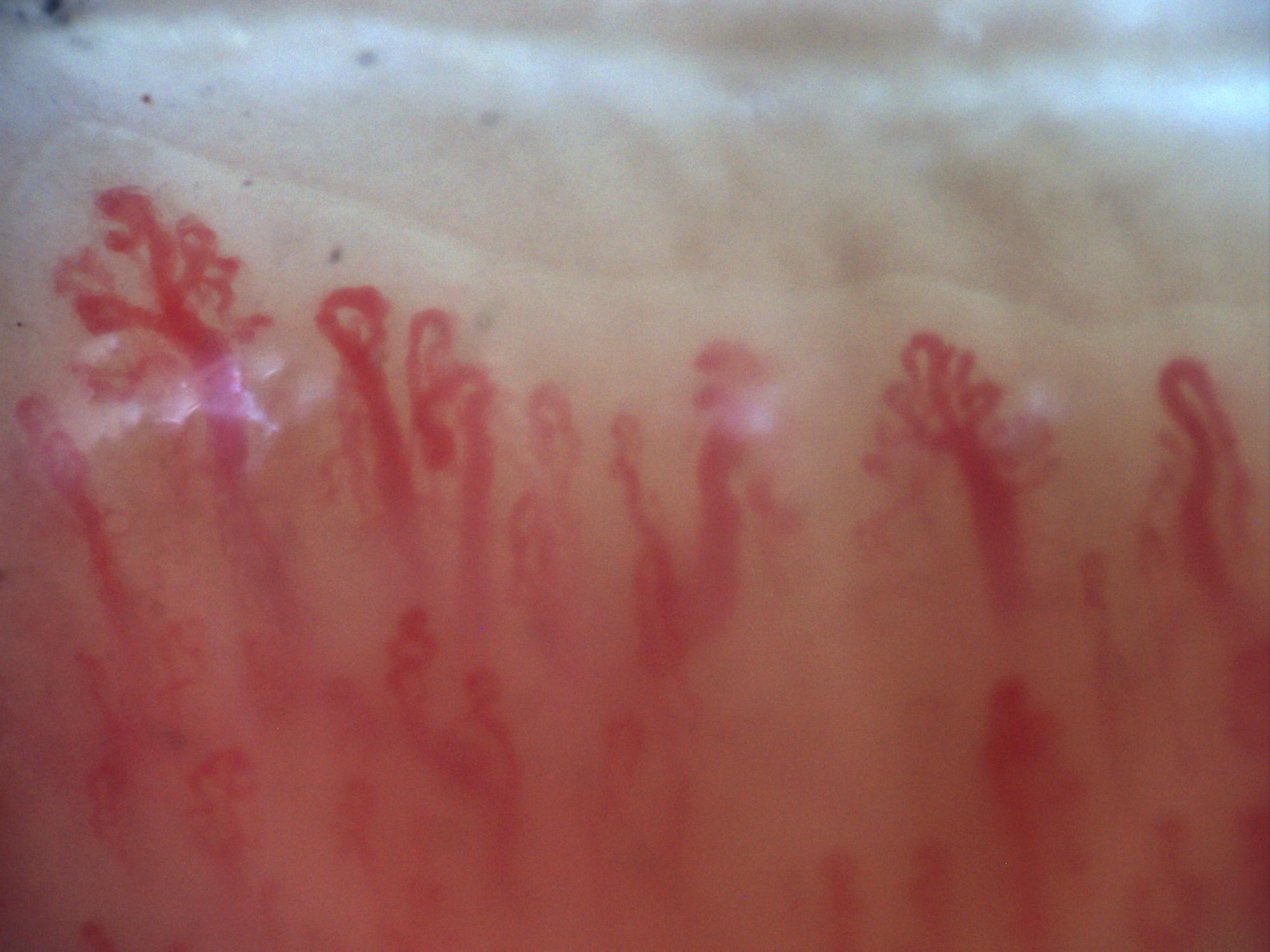 A. New vessel formation
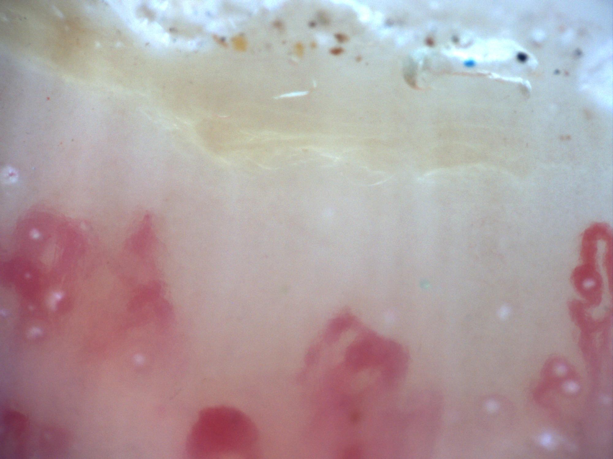 B. Avascular area
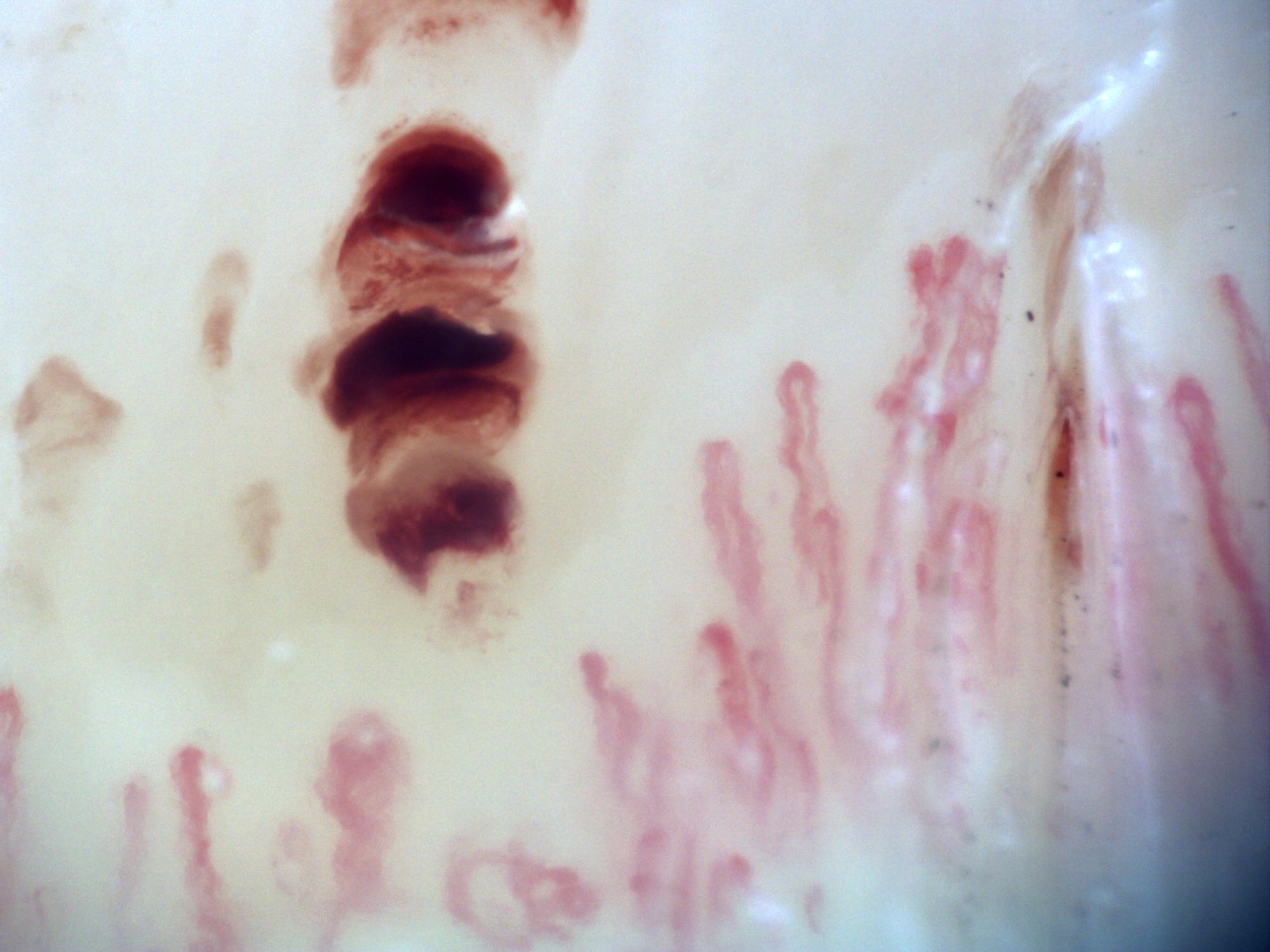 C. Hemorrhage
| Finding | Association with diabetes [OR (95% CI)] | Cautious interpretation |
|---|---|---|
| Tortuosity | 8.33 (2.31–30.04) | Frequent but nonspecific; carries more weight alongside other domains |
| Abnormal morphology | 5.00 (3.03–8.26) | Supports microvascular burden, but does not define a pattern on its own |
| Ectasia / dilation | 4.86 (2.56–9.20) | May reflect chronic vascular remodeling |
| Hemorrhages | 5.33 (2.31–12.34) | More relevant when multiple and not explained by trauma |
| Avascular areas | 6.14 (1.40–26.86) | Suggest rarefaction and may be associated with microvascular complications |
| Bushy capillaries / new vessel formation | 29.09* (5.90–143.55) | Strong association, although studies are few and results are not unanimous |
Results from a meta-analysis estimating the increased odds of microvascular abnormalities detectable by capillaroscopy in patients with diabetes [1].
*Bushy capillaries. CI, confidence interval; OR, odds ratio.
Retinopathy, nephropathy, and other complications
The area of greatest clinical interest is currently the relationship between capillaroscopy and established microvascular complications, particularly retinopathy and nephropathy [2, 3, 4, 7, 8, 9].
Diabetic retinopathy
One of the two 2025 meta-analyses cited above examined capillaroscopic findings in this complication. It included 1,349 patients with diabetes, 628 of whom had retinopathy. Patients with retinopathy consistently had larger arteriolar diameters and more bushy capillaries, new vessel formation, giant capillaries, tortuosity, hemorrhages, meandering capillaries, and avascular areas [2]. Some associations were substantial: the odds ratios (ORs) with 95% confidence intervals (CIs) were 8.37 (5.07–13.80) for giant capillaries and 7.92 (2.68–23.38) for avascular areas [2].
These findings align with earlier clinical studies. In 2018, Bakirci et al found more hemorrhages, ectasias, giant capillaries, and neoangiogenesis in patients with T2DM and retinopathy than in those without retinopathy [8]. In a later study, Okabe et al analyzed several variables longitudinally and observed progressive reductions in capillary number, length, and width with increasing retinopathy severity [9]. Together, these studies suggest a real association, but remain insufficient to replace fundus examination or standard ophthalmological screening techniques [2, 8, 9].
Diabetic nephropathy
The literature in this area has grown rapidly in recent years. A 2022 study found more capillary aneurysms, hemorrhages, tortuosity, new vessel formations, and bizarre capillaries in patients with albuminuria than in controls, and also found a correlation between albuminuria and tortuosity [3]. A recent larger cohort confirmed this finding, documenting significant differences according to eGFR and KDIGO categories, with more comma-shaped configurations and a higher proportion of abnormal shapes in groups with worse kidney function [4].
These observations do not establish capillaroscopy as a diagnostic test for retinopathy or nephropathy. They suggest that it may reflect part of the systemic microangiopathic burden and could eventually help improve multimodal risk models [2, 3, 4].

T1DM, T2DM, and prediabetes
Diabetes evidence does not all point in the same direction. It currently appears more robust in T2DM, while findings in T1DM have historically been less consistent [1, 7].
The 2025 meta-analysis found particularly pronounced associations in T2DM [1]. In a notable 2023 study, Lisco et al used computerized videocapillaroscopy in 102 patients with T2DM and observed an association between capillaroscopic abnormalities, clinical profile, and metabolic burden [7]. Earlier studies focused on T1DM did not always demonstrate clear morphological differences from controls, even with established retinopathy or vasculopathy [10].
Prediabetes also deserves attention as an intermediate setting. A study from the previous decade found more capillaroscopic distribution and morphological abnormalities in people with prediabetes than in healthy individuals, although the changes were less marked than in T2DM [11]. This suggests that early microvascular damage may be detectable at the nailfold, but evidence remains too limited to use the technique for population screening.

AI and datasets in diabetes
Diabetes is one of the settings in which combining capillaroscopy and artificial intelligence (AI) may offer the greatest potential, given the large number of patients and quantifiable microvascular outcomes [5].
A study recruiting 120 patients analyzed 5,236 images to develop a convolutional neural network model. Although its ability to predict cardiovascular events, retinopathy, albuminuria, or hypertension was modest, it achieved an area under the receiver operating characteristic curve (AUROC) of 0.84 for identifying diabetes [5]. This does not establish AI-assisted capillaroscopy as a clinical screening tool, but shows that nailfold images contain information that can be analyzed computationally.
Methodologically, diabetes offers both an advantage and a challenge. The potential data volume is enormous, allowing much larger datasets to be built and compared than in many rare rheumatic diseases. However, clinical labels are heterogeneous and depend on composite outcomes, disease duration, treatment, and comorbidities. As in other biomedical applications of AI, quality control and standardization are therefore as important as the algorithm itself [5].
Using the technique without overstating its capabilities
The main risk in diabetes is presenting capillaroscopy as an established screening test for retinopathy or nephropathy. The literature does not support that step [1, 2, 3, 4, 5].
- Use it as a complementary microangiopathy biomarker, not as a replacement for standard tests.
- Describe specific domains and avoid presenting a “diabetic pattern” as a validated construct.
- When relating findings to retinopathy or nephropathy, frame the conclusion as an association rather than a diagnosis.
- Prioritize T2DM with microvascular complications when selecting patients for research or exploratory assessment.
- When using AI, require image quality, prospective design, and external validation before inferring clinical utility.
Short cases
Case 1. T2DM with nonproliferative retinopathy
A patient with longstanding T2DM and retinopathy has marked tortuosity, hemorrhages, and avascular areas. Capillaroscopy is consistent with a high microvascular burden, but does not add more than fundus examination for retinopathy classification. Its value lies in supporting the systemic phenotype and, in cohort studies, improving risk models [2, 8, 9].
Case 2. A T2DM cohort with albuminuria
A research group analyzes tortuosity, abnormal shapes, and capillary density in patients with T2DM and different albuminuria levels. Capillaroscopy can quantify microangiopathy noninvasively and support subgroup comparisons, but interpretation still depends on eGFR, the albumin-to-creatinine ratio, and other conventional clinical variables [3, 4].
FAQ
Is there a specific capillaroscopic pattern for diabetes?
No validated specific pattern exists. It is more accurate to describe a set of frequent microvascular abnormalities than a unique pattern [1, 6].
Which type of diabetes shows the most consistent findings?
Mainly T2DM, where associations with capillaroscopic findings and microvascular complications are better documented [1, 2, 3, 4, 7].
Can capillaroscopy replace retinopathy screening?
No. Current evidence does not support using it instead of standard ophthalmological assessment [2, 8, 9].
Does it have value in diabetic nephropathy?
It may reflect microvascular burden and has been associated with albuminuria and worse kidney function, but does not replace standard kidney markers [3, 4].
Does AI change this field?
Yes, mainly in research and analysis of large datasets, but evidence remains insufficient to recommend routine clinical implementation [5].
Is it useful in prediabetes?
There are preliminary signs of early microvascular abnormalities, but clinical utility remains highly uncertain [11].
Glossary
- Diabetic microangiopathy
- Small-vessel damage associated with chronic hyperglycemia, endothelial inflammation, and vascular remodeling, affecting the retina, kidneys, peripheral nerves, and other tissues.
- Bushy capillary
- A capillary with an abnormal shape or marked branching, interpreted as a sign of neoangiogenesis or disorganized remodeling.
- Prediabetes
- A metabolic state preceding established diabetes, in which early signs of microvascular abnormalities may appear.
- KDIGO
- A chronic kidney risk classification system based on glomerular filtration rate and albuminuria.
- Research biomarker
- A variable useful for exploring associations or building prognostic models, even without an established clinical role.
References
- He W, Zhang M, Mao R, Han C, Shi L, Wu H. Association between nailfold capillaroscopic findings and diabetes: a systematic review and meta-analysis. Acta Diabetol. 2025;62(7):983-999. DOI: 10.1007/s00592-025-02520-4. PMID: 40332561.
- Yan Q, Chen SS, Lu XT, Chen H, Han CY, Wu HQ, et al. Association between findings in nailfold capillaroscopy and diabetic retinopathy: a systematic review and Meta-analysis. Int J Ophthalmol. 2025;18(6):1123-1130. DOI: 10.18240/ijo.2025.06.20. PMID: 40534817.
- Yilmaz U, Ayan A, Uyar S, Inci A, Ozer H, Yilmaz FT, et al. Capillaroscopic appearance of nailfold vasculature of diabetic nephropathy patients. Arch Endocrinol Metab. 2022;66(3):295-302. doi: 10.20945/2359-3997000000475. PMID: 35551680.
- Hsu PC, Liao PY, Huang SW, Chang HH, Chiang JY, Lo LC. Nailfold capillary abnormalities as indicators of diabetic nephropathy progression: a cross-sectional study in type 2 diabetes. Ann Med. 2025;57(1):2458766. DOI: 10.1080/07853890.2025.2458766. PMID: 39898556.
- Shah R, Petch J, Nelson W, Roth K, Noseworthy MD, Ghassemi M, et al. Nailfold capillaroscopy and deep learning in diabetes. J Diabetes. 2023;15(2):145-151. doi: 10.1111/1753-0407.13354. PMID: 36641812.
- Maldonado G, Guerrero R, Paredes C, Ríos C. Nailfold capillaroscopy in diabetes mellitus. Microvasc Res. 2017;112:41-46. doi: 10.1016/j.mvr.2017.03.001. PMID: 28274735.
- Lisco G, Triggiani V. Computerized nailfold video-capillaroscopy in type 2 diabetes: A cross-sectional study on 102 outpatients. J Diabetes. 2023;15(10):890-899. DOI: 10.1111/1753-0407.13442. PMID: 37424059.
- Bakirci S, Celik E, Acikgoz SB, Erturk Z, Tocoglu AG, Imga NN, et al. The evaluation of nailfold videocapillaroscopy findings in patients with type 2 diabetes with and without diabetic retinopathy. North Clin Istanb. 2018;6(2):146-150. doi: 10.14744/nci.2018.02222. PMID: 31297481.
- Okabe T, Kunikata H, Yasuda M, Kodama S, Maeda Y, Nakano J, et al. Relationship between nailfold capillaroscopy parameters and the severity of diabetic retinopathy. Graefes Arch Clin Exp Ophthalmol. 2024;262(3):759-768. doi: 10.1007/s00417-023-06220-z. PMID: 37874367.
- Trapp RG, Soler NG, Spencer-Green G. Nailfold capillaroscopy in type I diabetics with vasculopathy and limited joint mobility. J Rheumatol. 1986;13(5):917-920. PMID: 3820201.
- Hsu PC, Liao PY, Chang HH, Chiang JY, Huang YC, Lo LC. Nailfold capillary abnormalities are associated with type 2 diabetes progression and correlated with peripheral neuropathy. Medicine (Baltimore). 2016;95(52):e5714. doi: 10.1097/MD.0000000000005714. PMID: 28033273.