In antiphospholipid syndrome (APS) and vasculitis, capillaroscopy is far less validated than in systemic sclerosis or dermatomyositis. Current interest centers on two distinct settings: recognizing suggestive but nonspecific microvascular findings in APS, and exploring inflammatory burden or vascular damage in selected systemic vasculitides, particularly those associated with antineutrophil cytoplasmic antibodies (ANCA), where data linking findings to activity and organ involvement are beginning to emerge [1, 2, 3, 4, 5, 6, 7, 8, 9, 10, 11].
- There is no validated specific capillaroscopic pattern in APS; normal examinations or nonspecific abnormalities are usual [1, 2, 3, 4].
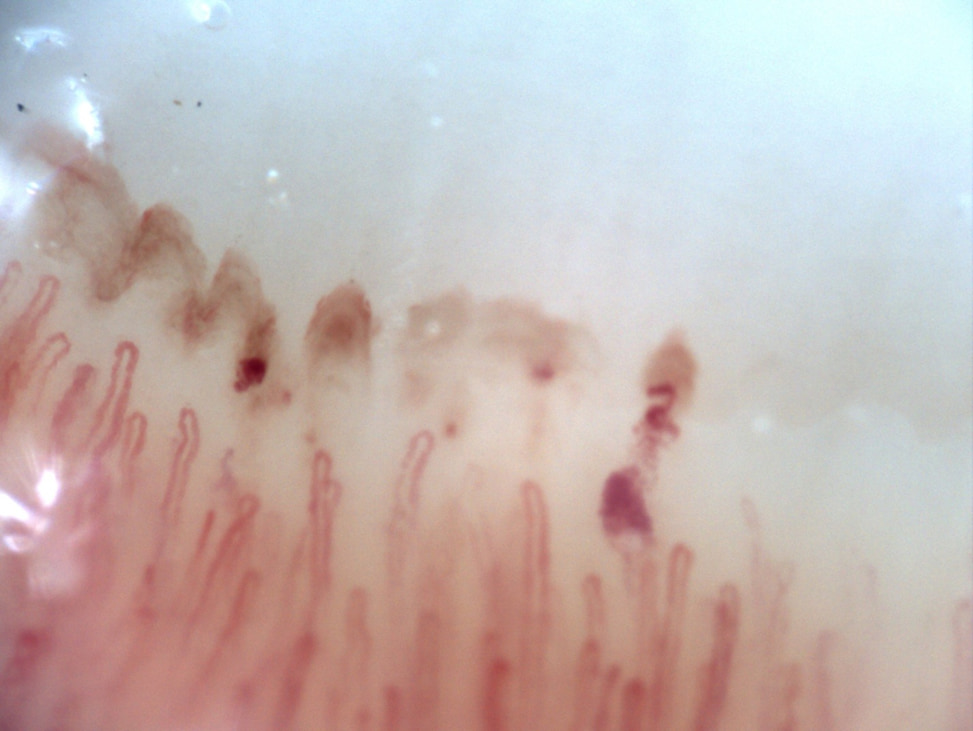
- Symmetrical hemorrhages are the most frequently cited classical finding in APS, but they are neither universal nor diagnostic, and may be confused with local trauma or the effects of anticoagulation [3, 4, 5, 6, 7].
- Comparison with anticoagulated controls matters: in Ferrari et al's study, hemorrhages were more frequent in APS than in antiphospholipid antibody carriers without the syndrome, but not more frequent than in controls anticoagulated for other reasons [3].
- In vasculitis, capillaroscopy appears more promising for assessing activity or microvascular damage than for classification; published findings remain exploratory [8, 9, 10, 11].
- In ANCA-associated vasculitis (AAV), hemorrhages, capillary loss, neoangiogenesis, and tortuosity have been associated with skin, lung, and kidney involvement, with partial improvement after immunosuppression [9].
- Cautious interpretation is essential: no capillary abnormality can replace APS criteria, antiphospholipid serology, biopsy, vascular imaging, or the standard immunological workup for vasculitis [1, 2, 3, 4, 5, 6, 7, 8, 9, 10, 11].
Learning path
If your clinical question concerns APS or vasculitis, this unit explains when capillaroscopy can support suspicion and when findings should not lead to excessive conclusions. The key is to avoid extrapolating the reasoning used in systemic sclerosis to these settings [1, 2, 3, 4, 5, 6, 7, 8, 9, 10, 11].
APS: what is established
The capillaroscopy literature on APS is scarce, fragmented, and based on small cohorts. This limitation shapes all subsequent clinical interpretation [1, 2, 3, 4, 5, 6, 7].
A large retrospective study of connective tissue diseases with Raynaud's phenomenon from a European Alliance of Associations for Rheumatology (EULAR) center showed that most patients with APS had normal or nonspecific capillaroscopy, with only a minority showing a pattern resembling scleroderma [2]. Ferrari et al reached a similar conclusion: both patients with APS and antiphospholipid antibody carriers without APS mainly had nonspecific abnormalities; the main difference was a greater frequency of hemorrhages in APS than in asymptomatic carriers [3].
The classical study by Vaz et al, focused on primary APS, added another qualification. It found morphological abnormalities in 77.8% of patients, but no clear functional reduction in density or conclusive disturbance of resting flow [4]. In practical terms, APS may produce visible microvascular injury, but this does not have a unique morphology or form an easily recognizable pattern.
Hemorrhages and interpretation pitfalls
Symmetrical hemorrhages are the most frequently cited abnormality in APS, but also the easiest to misinterpret when context is overlooked [3, 4, 5, 6, 7].
Sulli et al noted years ago that capillaroscopic abnormalities could be more frequent in patients with antiphospholipid antibodies [5]. Aslanidis et al went further, linking capillaroscopic hemorrhages with thrombosis and the antiphospholipid immunological profile in patients at high vascular risk [7]. However, these observations came from populations selected for high clinical suspicion and do not support a broadly applicable screening strategy.
Ferrari et al introduced a crucial methodological caution: when patients with APS are compared both with antibody carriers and with controls anticoagulated for non-immunological indications, the apparent discriminatory value of hemorrhages diminishes [3]. Before attributing hemorrhage to APS, consider two potential sources of error: local trauma, including cosmetic manipulation, and anticoagulation. Without this context, the finding loses much of its value.
| Finding | What it suggests | Main limitation |
|---|---|---|
| Symmetrical hemorrhages | May increase suspicion of clinical APS in a compatible setting [3, 5, 6, 7] | Nonspecific and potentially influenced by anticoagulation or trauma |
| Nonspecific pattern | The most frequent presentation [2, 3] | Adds little without a well-defined clinical question |
| Isolated scleroderma pattern | Reported, but uncommon [2] | Does not redefine the diagnosis or explain APS on its own |
Can capillaroscopy diagnose APS?
No, if diagnosis means replacing clinical and laboratory criteria. It may contribute in selected settings, but its performance is modest [1, 2, 3, 4, 5, 6, 7].
Pyrpasopoulou et al proposed capillaroscopy as a screening test for clinical APS, and Aslanidis et al incorporated it into a strategy for patients with signs of vasculopathy [6, 7]. These studies do not establish a sufficiently robust pattern to support a general recommendation. The most cautious current approach is that, in a patient with livedo, Raynaud's phenomenon, thrombosis, or a compatible obstetric history, symmetrical hemorrhages or marked microangiopathy may strengthen suspicion and prompt completion of the workup. They cannot establish the diagnosis without persistent serological positivity and the relevant criteria.
Conversely, normal capillaroscopy cannot exclude APS. The literature agrees that a substantial proportion of patients with confirmed APS have normal or only nonspecific examinations [2, 3]. This is crucial to avoid using the technique as a rule-out test.
Vasculitis: emerging evidence
Unlike APS research, the vasculitis literature focuses less on a typical finding and more on whether capillaroscopy can reflect microvascular inflammatory activity. This is an appealing but still preliminary hypothesis [8, 9, 10, 11].
Keret et al compared active systemic vasculitis, vasculitis in remission, and healthy controls, finding more neoangiogenesis and capillary loss in active disease. They also described two underexplored signs, rolling and peri-capillary stippling, potentially related to slowing of flow and capillary leakage [8]. These findings come from a small, heterogeneous cohort and generate hypotheses rather than establish new diagnostic terminology.
The strongest evidence so far comes from AAV. Among 51 patients, Matsuda et al found a microangiopathy pattern in 70.6%, a scleroderma pattern in 7.8%, and correlations between capillaroscopic scores and the severity of skin, lung, and kidney involvement. Hemorrhages also decreased after three months of immunosuppression [9]. Capillaroscopy could therefore serve as a complementary marker of activity and response in AAV, although multicenter validation is still needed.
More recently, Bhandare et al found that avascular areas, dropouts, receding capillaries, and disorganization were more frequent in cutaneous vasculitis than in controls, with more findings in patients with systemic involvement or ulcerated lesions [11]. This does not establish a specific pattern, but suggests the technique may detect microvascular burden in a subset of patients.
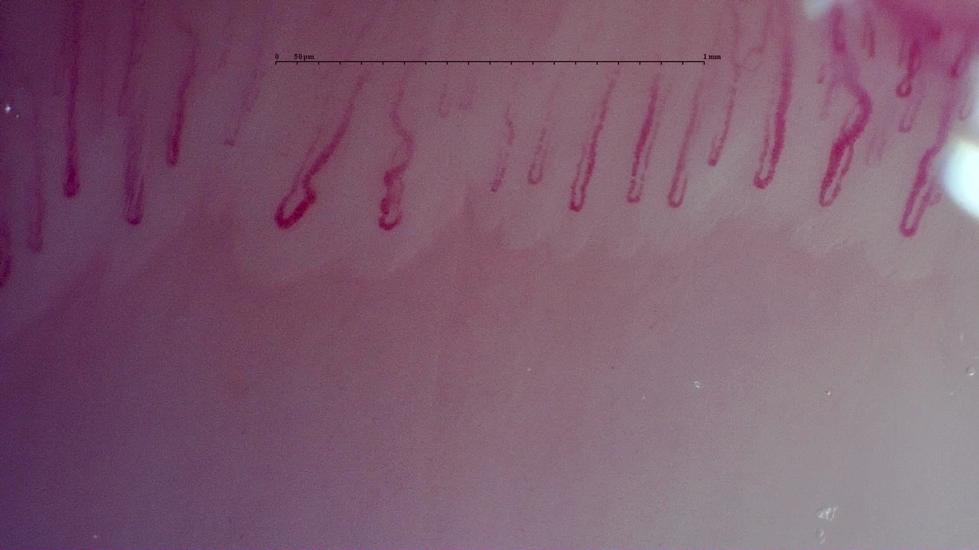 A. Capillary dilation · left middle finger (L3B)
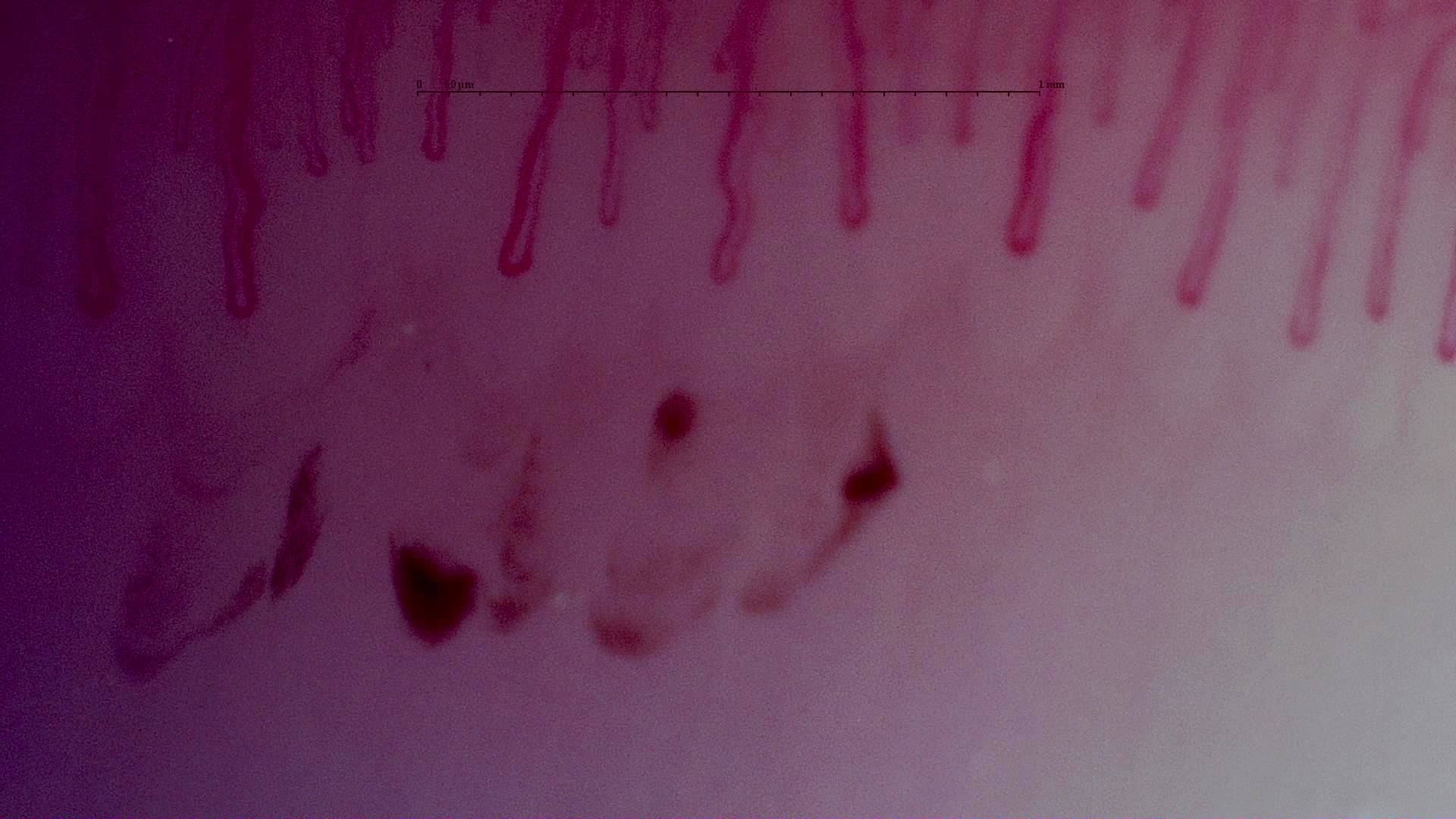 B. Hemorrhagic spots · right little finger (R5D)
AAV, Behçet's disease, and cutaneous vasculitis
Capillaroscopy does not have the same value in every vasculitis. It is useful to distinguish conditions with reproducible signals from those supported only by anecdotal findings [8, 9, 10, 11].
In AAV, the combination of hemorrhages, capillary loss, neoangiogenesis, and tortuosity appears consistent and is also associated with organ involvement [9]. In Behçet's disease, the Mercadé-Torras cohort found qualitative abnormalities, mainly tortuous or abnormal capillaries, in 47.7% of patients. Having two or more abnormalities was associated with vascular disease, particularly superficial thrombophlebitis [10]. These findings suggest possible value in identifying vascular phenotypes, but do not establish a specific Behçet's pattern.
In cutaneous vasculitis, the evidence is even more exploratory. A recent study using a handheld dermatoscope found more disorganization, dropouts, avascular areas, and receding capillaries in patients with systemic involvement, comorbidity, or ulcerated lesions [11]. This is clinically interesting because the technique is inexpensive and accessible, but the sample size and methods do not yet support a formal recommendation.
| Condition | Most frequently reported findings | Current contribution |
|---|---|---|
| APS | Symmetrical hemorrhages, nonspecific morphology [2, 3, 4, 5, 6, 7] | Contextual support in patients with high clinical suspicion |
| AAV | Hemorrhages, capillary loss, neoangiogenesis, tortuosity [9] | Possible complementary marker of activity and response |
| Behçet's disease | Tortuosity, abnormal shapes, isolated hemorrhages [10] | Exploratory signal of a vascular phenotype |
| Cutaneous vasculitis | Dropouts, avascular areas, receding capillaries [11] | Accessible complementary method for documenting microvascular burden |
Insufficient evidence / not found. There is insufficient published or practical support for using capillaroscopy as a universal screening tool for systemic vasculitis or as a replacement for biopsy, vascular imaging, or serological markers.
Practical reporting
In APS and vasculitis, a useful report should be even more cautious than in SLE. Overvaluing findings is a greater risk than underestimating them in these settings [1, 2, 3, 4, 5, 6, 7, 8, 9, 10, 11].
- Describe whether hemorrhages are symmetrical, focal, extensive, or compatible with trauma.
- If the patient receives anticoagulation, explicitly include this in the interpretation.
- In vasculitis, prioritize domains such as capillary loss, neoangiogenesis, tortuosity, and disorganization before assigning a pattern.
- Avoid terms such as “capillaroscopy diagnostic of APS” or “vasculitis confirmed by the image.”
- Use conditional conclusions: “findings compatible with nonspecific microangiopathy” or “abnormalities that may accompany vascular activity.”
Short cases
Case 1. Anticoagulated APS with hemorrhages
A woman with thrombotic APS receiving warfarin has symmetrical nailfold hemorrhages. The finding is compatible with the syndrome and may support the vascular context, but must be interpreted alongside anticoagulant treatment. It does not change the diagnosis or treatment strategy on its own [3, 4, 5, 6, 7].
Case 2. AAV with lung and kidney involvement
A patient with active AAV has capillary loss, hemorrhages, and neoangiogenesis. Capillaroscopy adds a signal of microvascular activity consistent with the systemic picture and may help document response after immunosuppression. Treatment decisions still depend on the Birmingham Vasculitis Activity Score (BVAS), kidney function, imaging, and histology when appropriate [8, 9].
FAQ
Is there a specific APS pattern?
No. Symmetrical hemorrhages are the most frequently cited finding, but do not constitute a validated specific pattern [2, 3, 4, 5, 6, 7].
Can capillaroscopy diagnose APS?
No. It can support suspicion in an appropriate setting, but does not replace clinical criteria or persistent serological positivity [1, 2, 3, 4, 5, 6, 7].
Can anticoagulation complicate interpretation?
Yes. It is one of the main confounders when interpreting hemorrhages in APS [3].
Which vasculitis has the most consistent evidence so far?
AAV has shown the clearest associations between capillaroscopic findings and clinical severity [9].
Glossary
- APS
- Antiphospholipid syndrome, a thromboinflammatory condition defined by clinical criteria and persistently positive serological findings.
- Symmetrical hemorrhages
- Repeated capillary extravasations across several fingers in a relatively regular distribution; suggestive in APS, but nonspecific.
- Rolling
- An exploratory term proposed in systemic vasculitis for visible slowing of capillary flow.
- Peri-capillary stippling
- Punctate pericapillary deposits described in a small cohort with active vasculitis, still lacking broad external validation.
- AAV
- ANCA-associated vasculitis, a group of small-vessel vasculitides in which capillaroscopy has shown greater clinical utility outside the scleroderma spectrum.
References
- Smith V, Ickinger C, Hysa E, Snow M, Frech T, Sulli A, et al. Nailfold capillaroscopy. Best Pract Res Clin Rheumatol. 2023;37(1):101849. doi: 10.1016/j.berh.2023.101849. PMID: 37419757.
- Hysa E, Pizzorni C, Sammorì S, Gotelli E, Cere A, Schenone C, et al. Microvascular damage in autoimmune connective tissue diseases: a capillaroscopic analysis from 20 years of experience in a EULAR training and research referral centre for imaging. RMD Open. 2023;9(3):e003071. DOI: 10.1136/rmdopen-2023-003071. PMID: 37451812.
- Ferrari G, Gotelli E, Paolino S, Pesce G, Nanni L, Colombo BM, et al. Antiphospholipid antibodies and anticoagulant therapy: capillaroscopic findings. Arthritis Res Ther. 2021;23(1):175. doi: 10.1186/s13075-021-02551-6. PMID: 34176504.
- Vaz JL, Dancour MA, Bottino DA, Bouskela E. Nailfold videocapillaroscopy in primary antiphospholipid syndrome (PAPS). Rheumatology (Oxford). 2004;43(8):1025-7. doi: 10.1093/rheumatology/keh233. PMID: 15187240.
- Sulli A, Pizzorni C, Cutolo M. Nailfold videocapillaroscopy abnormalities in patients with antiphospholipid antibodies. J Rheumatol. 2000;27(6):1574-1576. PMID: 10852305.
- Pyrpasopoulou A, Triantafyllou A, Anyfanti P, Douma S, Aslanidis S. Capillaroscopy as a screening test for clinical antiphospholipid syndrome. Eur J Intern Med. 2011;22(6):e158-e159. DOI: 10.1016/j.ejim.2011.07.007. PMID: 22075311.
- Aslanidis S, Pyrpasopoulou A, Doumas M, Triantafyllou A, Chatzimichailidou S, Zamboulis C. Association of capillaroscopic microhaemorrhages with clinical and immunological antiphospholipid syndrome. Clin Exp Rheumatol. 2011;29(2):307-309. PMID: 21385543.
- Keret S, Mazzawi J, Slobodin G, Rimar O, Rosner I, Rozenbaum M, et al. Nailfold video capillaroscopy as a useful diagnostic tool in systemic vasculitis. Microvasc Res. 2022;143:104406. DOI: 10.1016/j.mvr.2022.104406. PMID: 35835173.
- Matsuda S, Kotani T, Wakura R, Suzuka T, Kuwabara H, Kiboshi T, et al. Examination of nailfold videocapillaroscopy findings in ANCA-associated vasculitis. Rheumatology (Oxford). 2023;62(2):747-757. DOI: 10.1093/rheumatology/keac402. PMID: 35816001.
- Mercadé-Torras JM, Guillén-Del-Castillo A, Buján S, Solans-Laque R. Nailfold videocapillaroscopy abnormalities and vascular manifestations in Behçet's syndrome. Clin Exp Rheumatol. 2024;42(10):2065-2070. DOI: 10.55563/clinexprheumatol/v5mz8d. PMID: 39404484.
- Bhandare P, Naik R, Mahesh Y, Ghodge R. Nailfold Capillaroscopic (NFC) Changes in Vasculitis and Their Correlation with Systemic Involvement: A Cross-Sectional Study. Indian Dermatol Online J. 2025;16(6):928-933. DOI: 10.4103/idoj.idoj_859_24. PMID: 40814970.