Fundamentals of capillaroscopy
Core conceptual and clinical foundations for rigorous interpretation of nailfold capillaroscopy.
Overview
Modules and units
Choose a module and jump straight into the unit you need. Each block is ordered so you can keep context without reading a long roadmap first.
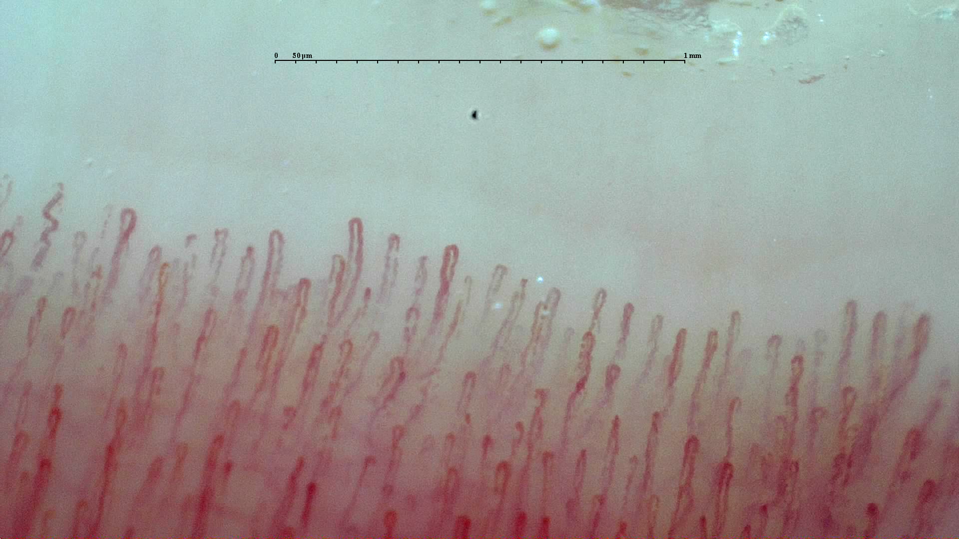
Module 1
Foundations
What capillaroscopy is, why the periungual region matters, and which anatomy and classical reading frameworks you need before interpreting anything.
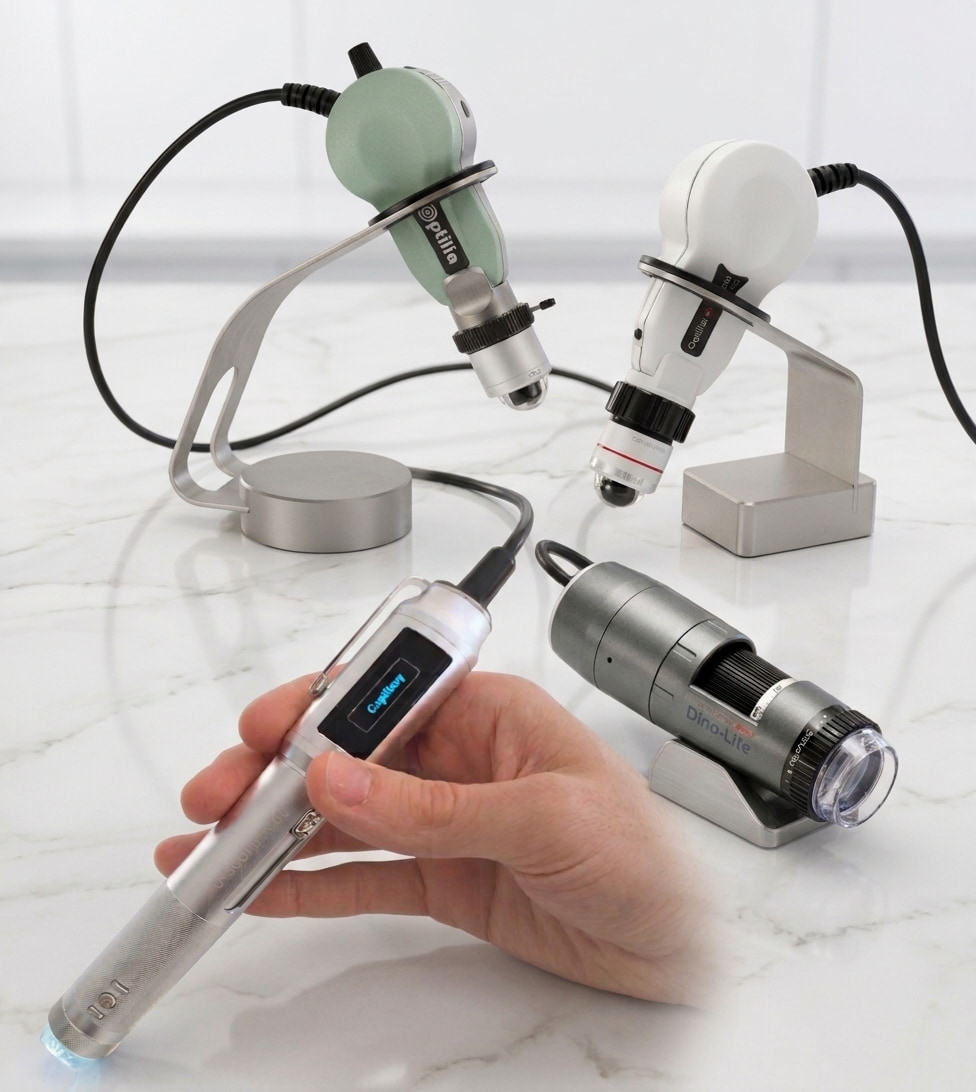
Module 2
Technique and workflow
Equipment, acquisition, quality control, quantification, and AI. This block gathers what you need for usable and comparable studies.
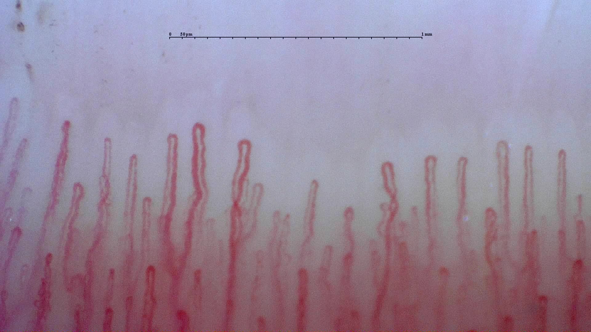
Module 3
Interpretation
Normality, variability, descriptive language, and reporting. This is where you learn not to overcall and how to write useful conclusions.
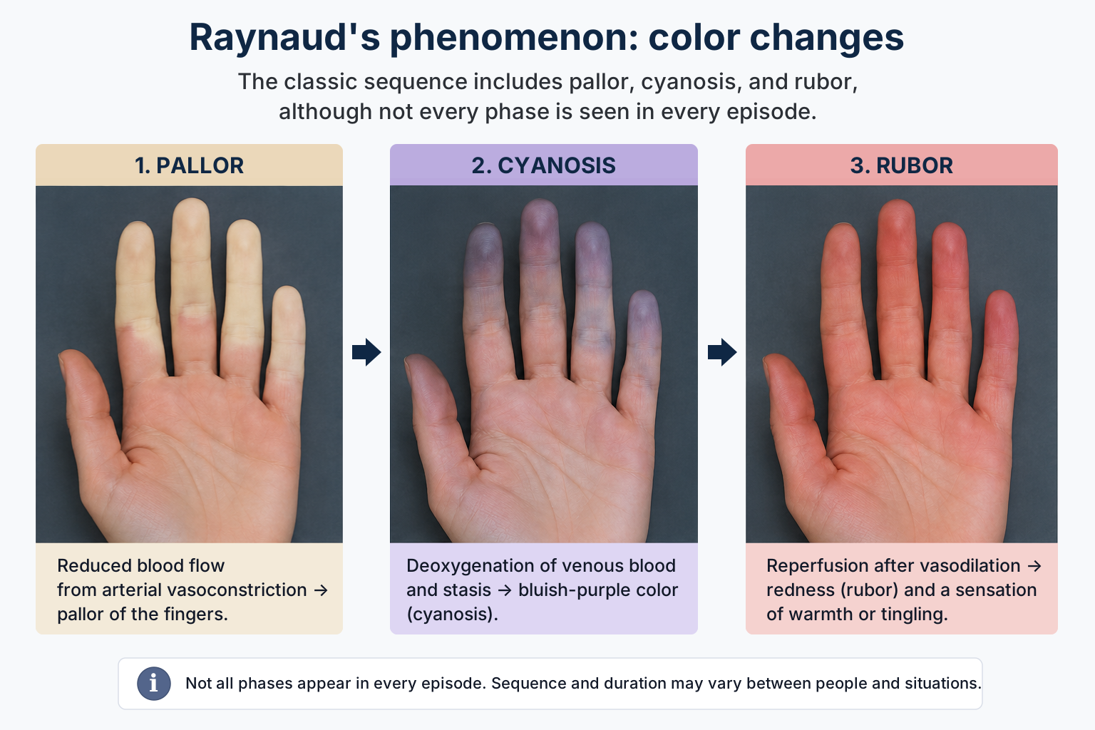
Module 4
Initial clinical application
When to request the test, how to use it in Raynaud's phenomenon, and how to distinguish it from other acrosyndromes before disease-specific chapters.
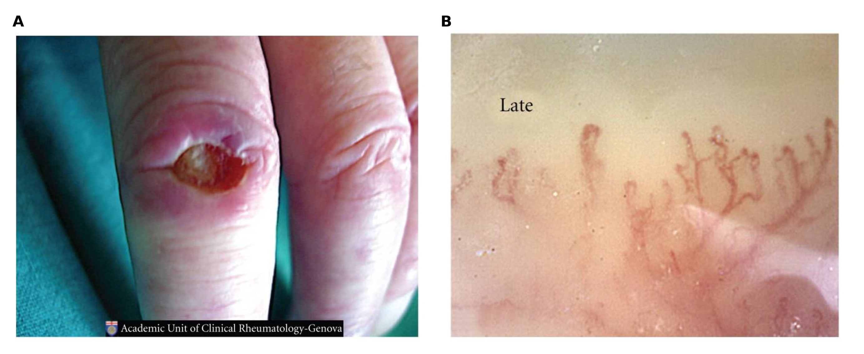
Module 5
Systemic sclerosis
A dedicated block for the scleroderma pattern, classification, vascular prognosis, and longitudinal follow-up.
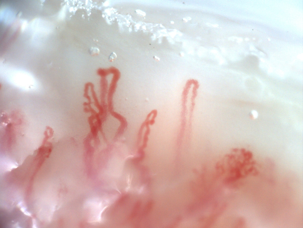
Module 6
Other autoimmune diseases
Overlap disease, inflammatory myopathies, lupus, APS, and vasculitis, where capillaroscopy mainly contributes phenotype context and stratification.
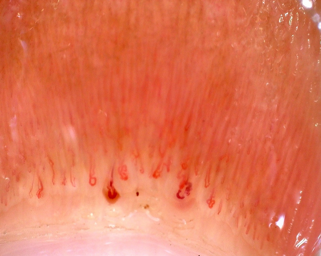
Module 7
Transversal extensions
Units that expand the map beyond the central pathway, such as pediatrics or diabetes, without changing the core learning sequence.
Show framework references
- Smith V, et al. Nailfold capillaroscopy. Best Pract Res Clin Rheumatol. 2023;37(1):101849.
- Smith V, et al. Standardisation of nailfold capillaroscopy for the assessment of patients with Raynaud's phenomenon and systemic sclerosis. Autoimmun Rev. 2020;19(3):102458.
- Bellando-Randone S, et al. Progression of patients with Raynaud's phenomenon to systemic sclerosis: VEDOSS registry. Lancet Rheumatol. 2021;3(12):e834-e843.